Reproductive System A&P
Shared 4/1/2026•66 views
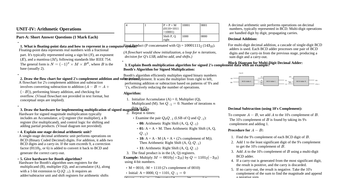
### Male Reproductive Anatomy - **Scrotum:** Pouch of skin and superficial fascia housing the testes. - **Dartos muscle:** Smooth muscle, wrinkles scrotal skin, pulls testes close to body. - **Cremaster muscle:** Skeletal muscle, elevates testes. - **Temperature regulation:** Keeps testes 2-3°C cooler than body for spermatogenesis. - **Testes (Gonads):** Produce sperm and testosterone. - **Tunica vaginalis:** Outer serous layer from peritoneum. - **Tunica albuginea:** Inner fibrous capsule. - **Seminiferous tubules:** Site of spermatogenesis. - **Spermatogenic cells:** Form sperm. - **Sustentocytes (Sertoli cells):** Support, nourish developing sperm, form blood-testis barrier. - **Interstitial endocrine cells (Leydig cells):** Produce androgens (e.g., testosterone) in connective tissue surrounding seminiferous tubules. - **Epididymis:** Coiled tube on posterior side of testis. - Stores and matures sperm (takes ~20 days). - Sperm gain motility here. - **Ductus Deferens (Vas Deferens):** Transports sperm from epididymis to ejaculatory duct. - Part of the spermatic cord. - Ampulla of ductus deferens joins seminal vesicle duct to form ejaculatory duct. - **Ejaculatory Duct:** Formed by union of ductus deferens and seminal vesicle duct; passes through prostate. - **Urethra:** Extends from bladder to tip of penis; three regions: - **Prostatic urethra:** Through prostate. - **Membranous urethra:** Through urogenital diaphragm. - **Spongy (penile) urethra:** Through penis. - **Accessory Glands:** Produce seminal fluid. - **Seminal Vesicles (2):** Posterior to bladder. Produce viscous alkaline fluid (60-70% of semen volume) containing fructose, citric acid, coagulating enzyme (vesiculase), and prostaglandins. - **Prostate Gland (1):** Inferior to bladder, encircles urethra. Produces milky, slightly acidic fluid (20-30% of semen volume) containing citrate, enzymes (e.g., prostate-specific antigen, PSA), and seminalplasmin (antibiotic). - **Bulbo-urethral Glands (Cowper's Glands) (2):** Inferior to prostate. Produce thick, clear mucus that lubricates glans penis and neutralizes acidic urine in urethra before ejaculation. - **Penis:** Male copulatory organ. - **Root:** Attached portion. - **Shaft (Body):** Contains erectile tissue. - **Glans Penis:** Enlarged tip. - **Prepuce (Foreskin):** Loose skin covering glans (removed during circumcision). - **Corpora Cavernosa (2):** Paired dorsal erectile bodies. - **Corpus Spongiosum (1):** Surrounds urethra, expands to form glans and bulb of penis. ### Male Reproductive Physiology - **Spermatogenesis:** Production of sperm in seminiferous tubules. - **Spermatogonia:** Stem cells (diploid, 2n), undergo mitosis. - **Primary spermatocytes:** Undergo Meiosis I to form two secondary spermatocytes (haploid, n). - **Secondary spermatocytes:** Undergo Meiosis II to form two spermatids (haploid, n). - **Spermiogenesis:** Spermatids mature into spermatozoa (sperm). - **Sperm structure:** - **Head:** Contains nucleus (DNA) and acrosome (enzymes for egg penetration). - **Midpiece:** Contains mitochondria (ATP for motility). - **Tail (Flagellum):** For propulsion. - **Hormonal Regulation:** - **GnRH (Gonadotropin-releasing hormone):** From hypothalamus, stimulates anterior pituitary. - **FSH (Follicle-stimulating hormone):** From anterior pituitary, stimulates sustentocytes to release androgen-binding protein (ABP), enhancing testosterone's effect on spermatogenesis. - **LH (Luteinizing hormone):** From anterior pituitary, stimulates interstitial endocrine cells to secrete testosterone. - **Testosterone:** Produced by Leydig cells. - Stimulates spermatogenesis. - Promotes development of secondary sex characteristics (deep voice, body hair, muscle mass). - Boosts libido. - **Inhibin:** Produced by sustentocytes, inhibits FSH release when sperm count is high. - **Erection:** Parasympathetic reflex. Nitric oxide (NO) release causes vasodilation of arterioles supplying erectile tissue, filling spaces with blood. - **Ejaculation:** Sympathetic reflex. - Ducts and glands contract, emptying contents. - Bladder sphincter constricts. - Bulbospongiosus muscles contract rhythmically. ### Female Reproductive Anatomy - **Ovaries (Gonads):** Produce ova (eggs), estrogen, and progesterone. - **Tunica albuginea:** Outer fibrous capsule. - **Cortex:** Outer region, contains ovarian follicles. - **Medulla:** Inner region, contains blood vessels and nerves. - **Ovarian follicles:** Immature egg (oocyte) surrounded by follicle cells. - **Primordial follicle:** Single layer of squamous follicle cells. - **Primary follicle:** Single layer of cuboidal follicle cells. - **Secondary follicle:** Multiple layers of granulosa cells. - **Vesicular (Graafian) follicle:** Mature follicle with antrum (fluid-filled cavity). - **Corpus luteum:** Remnant of ruptured follicle after ovulation; produces progesterone and some estrogen. - **Corpus albicans:** Degenerated corpus luteum. - **Uterine Tubes (Fallopian Tubes/Oviducts):** Receive ovulated oocyte; site of fertilization. - **Infundibulum:** Funnel-shaped end with fimbriae (ciliated projections) that sweep oocyte into tube. - **Ampulla:** Usual site of fertilization. - **Isthmus:** Narrowed region joining uterus. - Cilia and muscular peristalsis move oocyte towards uterus. - **Uterus:** Receives, retains, and nourishes fertilized ovum. - **Fundus:** Rounded superior region. - **Body (Corpus):** Main portion. - **Isthmus:** Narrow constriction. - **Cervix:** Narrow neck projecting into vagina. - **Cervical canal:** Connects uterus to vagina. - **Internal os:** Opening into uterus. - **External os:** Opening into vagina. - **Uterine Wall Layers:** - **Perimetrium:** Outermost serous layer (visceral peritoneum). - **Myometrium:** Middle layer of smooth muscle; contracts during childbirth. - **Endometrium:** Inner mucosal lining; site of implantation. - **Stratum functionalis (functional layer):** Shed during menstruation. - **Stratum basalis (basal layer):** Forms new functionalis after menstruation. - **Vagina:** Female copulatory organ; birth canal; passageway for menstrual flow. - Extends from cervix to exterior. - **Hymen:** Incomplete partition near vaginal orifice. - **External Genitalia (Vulva/Pudendum):** - **Mons Pubis:** Fatty area overlying pubic symphysis. - **Labia Majora:** Outer folds, homologous to scrotum. - **Labia Minora:** Inner folds. - **Clitoris:** Erectile tissue, homologous to penis. - **Glans clitoris:** Exposed portion. - **Prepuce of clitoris:** Hood. - **Vestibule:** Area enclosed by labia minora; contains vaginal and urethral openings. - **Greater Vestibular Glands (Bartholin's Glands):** Flank vaginal opening; secrete mucus for lubrication. ### Female Reproductive Physiology - **Oogenesis:** Production of female gametes (ova) in ovaries. - **Oogonia:** Stem cells (diploid, 2n) in fetal ovary, undergo mitosis. - **Primary oocytes:** Begin Meiosis I in fetal period, arrest in Prophase I. - At puberty, one primary oocyte per month completes Meiosis I, forming a large **secondary oocyte** (haploid, n) and a tiny first polar body. - Secondary oocyte arrests in Metaphase II; ovulated. - If fertilized, secondary oocyte completes Meiosis II, forming a large ovum and a second polar body. - **Ovarian Cycle:** Monthly series of events associated with maturation of an egg. - **Follicular Phase (Day 1-14):** Follicle growth and estrogen secretion. - Primordial follicle → Primary → Secondary → Vesicular follicle. - FSH stimulates follicle growth. - Granulosa cells produce estrogen. - **Ovulation (Day 14):** Rupture of vesicular follicle and release of secondary oocyte. - Triggered by a surge in LH (due to high estrogen levels). - **Luteal Phase (Day 14-28):** Formation and degeneration of corpus luteum. - Ruptured follicle transforms into corpus luteum under LH influence. - Corpus luteum produces large amounts of progesterone and some estrogen. - If no pregnancy, corpus luteum degenerates into corpus albicans (~10 days), progesterone and estrogen levels drop. - **Uterine (Menstrual) Cycle:** Monthly changes in endometrial lining in response to ovarian hormones. - **Menstrual Phase (Day 1-5):** Shedding of stratum functionalis. - Low ovarian hormones cause endometrial arteries to constrict, tissue dies and is shed. - **Proliferative Phase (Day 6-14):** Endometrium rebuilds. - Rising estrogen (from growing follicles) stimulates regeneration of stratum functionalis. - Glands enlarge, arteries proliferate. - **Secretory Phase (Day 15-28):** Endometrium prepares for implantation. - Rising progesterone (from corpus luteum) enhances endometrial vascularization and gland secretion (glycogen, nutrients). - If no fertilization, corpus luteum degenerates, hormone levels drop, cycle restarts. - **Hormonal Regulation:** - **GnRH:** From hypothalamus, stimulates anterior pituitary. - **FSH:** From anterior pituitary, stimulates follicle growth and estrogen production. - **LH:** From anterior pituitary, triggers ovulation and corpus luteum formation/maintenance. - **Estrogen:** Produced by follicles. - Promotes oogenesis and follicle growth. - Induces secondary sex characteristics (breast development, widened hips). - During proliferative phase, rebuilds endometrium. - High levels trigger LH surge. - **Progesterone:** Produced by corpus luteum. - Maintains secretory endometrium for pregnancy. - Inhibits uterine motility. - Helps maintain pregnancy. - **Inhibin:** Produced by granulosa cells, inhibits FSH release. - **Mammary Glands:** Modified sweat glands that produce milk. - **Areola:** Pigmented skin surrounding nipple. - **Lactiferous ducts:** Carry milk to nipple. - **Lactiferous sinuses:** Store milk. ### Pregnancy & Development - **Fertilization:** Fusion of sperm and egg, typically in ampulla of uterine tube. - Zygote formed (diploid, 2n). - **Cleavage:** Rapid mitotic divisions of zygote. - **Morula:** Solid ball of 16+ cells. - **Blastocyst:** Hollow ball of cells with inner cell mass (embryoblast) and outer trophoblast. - **Implantation:** Blastocyst burrows into endometrium (~6-9 days post-fertilization). - **Trophoblast cells:** Secrete hCG (human chorionic gonadotropin), which maintains corpus luteum. - **Placenta:** Formed from embryonic (chorion) and maternal (decidua basalis) tissues. - Functions: Nutrient/gas/waste exchange, hormone production (hCG, estrogen, progesterone). - **Gestation:** ~280 days from last menstrual period. - **First Trimester:** Organogenesis, most susceptible to teratogens. - **Second Trimester:** Growth, maturation, fetal movements felt. - **Third Trimester:** Rapid growth, organ systems mature for survival outside womb. - **Childbirth (Parturition):** - **Initiation:** Fetal cortisol and maternal oxytocin (from posterior pituitary) play key roles. - **Stages of Labor:** 1. **Dilation Stage:** Cervix dilates (up to 10 cm), effacement (thinning) occurs. Longest stage. 2. **Expulsion Stage:** Fetus delivered. 3. **Placental Stage:** Placenta delivered (afterbirth). - **Lactation:** Milk production by mammary glands. - **Prolactin:** From anterior pituitary, stimulates milk production. - **Oxytocin:** From posterior pituitary, stimulates milk ejection (let-down reflex).