Embryology Essentials
Cheatsheet Content
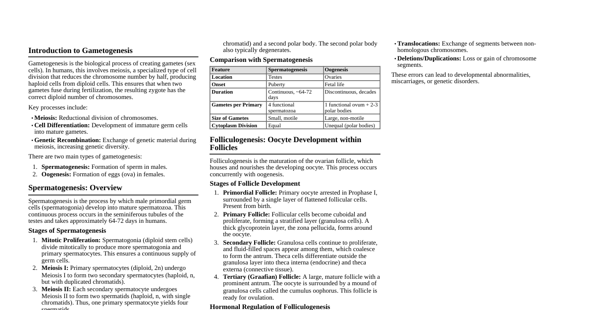
1. Gametogenesis Spermatogenesis Process: Spermatogonia $\rightarrow$ Primary spermatocytes $\rightarrow$ Secondary spermatocytes $\rightarrow$ Spermatids $\rightarrow$ Spermatozoa Location: Seminiferous tubules of testes Timing: Begins at puberty, continuous Meiosis I: Primary spermatocyte (diploid) $\rightarrow$ 2 Secondary spermatocytes (haploid) Meiosis II: Secondary spermatocyte (haploid) $\rightarrow$ 2 Spermatids (haploid) Spermiogenesis: Maturation of spermatids into spermatozoa (loss of cytoplasm, acrosome formation, flagellum development) Oogenesis Process: Oogonia $\rightarrow$ Primary oocytes $\rightarrow$ Secondary oocyte + First polar body $\rightarrow$ Ovum + Second polar body Location: Ovaries Timing: Fetal life: Oogonia proliferate, differentiate into primary oocytes, enter Meiosis I (prophase I arrest). Puberty: Monthly, one primary oocyte completes Meiosis I, forming secondary oocyte and first polar body. Ovulation: Secondary oocyte arrested in Meiosis II (metaphase II arrest). Fertilization: Meiosis II completes, forming ovum and second polar body. 2. Fertilization Definition: Fusion of male and female gametes. Location: Ampulla of uterine tube. Steps: Penetration of corona radiata (hyaluronidase from sperm). Penetration of zona pellucida (acrosin from acrosome reaction). Fusion of sperm and oocyte cell membranes. Cortical reaction: Release of cortical granules, zona pellucida hardens (block to polyspermy). Completion of Meiosis II by oocyte. Formation of male and female pronuclei. Fusion of pronuclei $\rightarrow$ Zygote (diploid). 3. Cleavage & Blastulation Cleavage Rapid mitotic divisions of zygote without significant growth. Increases cell number (blastomeres). Occurs as zygote moves down uterine tube. Morula: Ball of 12-32 blastomeres (approx. Day 3-4). Blastulation Morula enters uterus, fluid penetrates, forming blastocyst cavity (blastocele). Blastocyst: Inner Cell Mass (ICM) / Embryoblast: Forms embryo proper. Outer Cell Mass (OCM) / Trophoblast: Forms placenta. Zona pellucida degenerates (hatching of blastocyst) around Day 5-6, allowing implantation. 4. Implantation Definition: Blastocyst embeds in uterine endometrium. Timing: Begins Day 6-7, completed by Day 10-12. Location: Usually posterior wall of uterus. Trophoblast differentiates: Cytotrophoblast: Inner layer, mitotically active. Syncytiotrophoblast: Outer multinucleated mass, invades endometrium, produces hCG. Decidual reaction: Endometrial cells swell, accumulate glycogen and lipids. 5. Bilaminar Germ Disc (Week 2) Embryoblast differentiates into two layers: Epiblast: Dorsal layer, columnar cells. Forms floor of amniotic cavity. Hypoblast: Ventral layer, cuboidal cells. Forms roof of primary umbilical vesicle (yolk sac). Amniotic cavity and primary umbilical vesicle (yolk sac) form. Extraembryonic mesoderm forms, surrounding amnion and yolk sac. Chorionic cavity (extraembryonic coelom) forms within extraembryonic mesoderm. Connecting stalk: Attaches bilaminar disc to trophoblast, precursor to umbilical cord. 6. Gastrulation (Week 3) Formation of three germ layers from epiblast. Key event: Primitive streak appears on epiblast. Cells from epiblast migrate through primitive streak: Some displace hypoblast $\rightarrow$ Endoderm. Some form layer between epiblast and endoderm $\rightarrow$ Mesoderm. Remaining epiblast cells $\rightarrow$ Ectoderm. Notochord: Forms from mesoderm cells migrating through primitive node, induces neural plate. Germ Layer Derivatives Ectoderm: Surface Ectoderm: Epidermis, hair, nails, glands, tooth enamel, lens of eye, anterior pituitary. Neuroectoderm (Neural Tube): CNS (brain, spinal cord), retina, posterior pituitary. Neural Crest: PNS ganglia, Schwann cells, adrenal medulla, melanocytes, facial skeleton, odontoblasts. Mesoderm: Paraxial Mesoderm: Somites (sclerotome $\rightarrow$ vertebrae/ribs; myotome $\rightarrow$ skeletal muscle; dermatome $\rightarrow$ dermis). Intermediate Mesoderm: Urogenital system (kidneys, gonads, ducts). Lateral Plate Mesoderm: Somatic (parietal) layer: Limbs, body wall. Splanchnic (visceral) layer: Smooth muscle of gut, serous membranes, heart, blood vessels. Head Mesoderm: Connective tissue of head, eye muscles. Endoderm: GI tract epithelium, liver, pancreas. Respiratory tract epithelium (trachea, bronchi, lungs). Thyroid, parathyroid, thymus. Bladder, urethra epithelium. Eustachian tube, tonsils. 7. Neurulation (Weeks 3-4) Formation of neural tube from neural plate. Notochord induces overlying ectoderm to thicken $\rightarrow$ Neural plate. Neural plate folds $\rightarrow$ Neural groove and neural folds. Neural folds fuse $\rightarrow$ Neural tube. Neural crest cells delaminate from neural folds. Neural tube closes first in cervical region, then proceeds cranially and caudally. Anterior neuropore closes by Day 25, posterior neuropore by Day 28. Failure of closure $\rightarrow$ Neural tube defects (anencephaly, spina bifida). 8. Folding of Embryo (Week 4) Embryo folds in two planes: Craniocaudal (cephalocaudal) folding: Head and tail folds due to rapid growth of neural tube. Lateral folding: Lateral body wall folds towards midline. Results in: Formation of definitive gut tube (foregut, midgut, hindgut). Incorporation of part of yolk sac into gut. Formation of body cavities. Ventrolateral body wall closure. 9. Fetal Period (Week 9 - Birth) Primarily growth and maturation of tissues and organs. Less susceptible to teratogens compared to embryonic period (weeks 3-8), but functional defects possible. Key developments: Month 3: Head growth slows, ossification centers appear, external genitalia distinguishable. Month 4-5: Rapid growth in length, fetal movements ("quickening"). Month 6-7: Weight gain, lungs develop capacity for gas exchange. Month 8-9: Subcutaneous fat deposition, maturation of lungs. 10. Placenta Functions: Gas exchange, nutrient exchange, waste removal, hormone production (hCG, progesterone, estrogen). Components: Fetal part: Chorion frondosum (villous chorion). Maternal part: Decidua basalis. Structure: Chorionic villi project into intervillous spaces filled with maternal blood. Placental barrier: Syncytiotrophoblast, cytotrophoblast, connective tissue, fetal capillary endothelium. 11. Amniotic Fluid Origin: Initially from maternal blood, later fetal urine, secretions from respiratory tract. Functions: Protection (shock absorber), temperature regulation, fetal movement, lung development. Volume: ~$800-1000$ mL at term. Disorders: Polyhydramnios: Excess fluid ($>2000$ mL), often associated with esophageal atresia, anencephaly. Oligohydramnios: Low fluid ($ 12. Common Congenital Anomalies Neural Tube Defects: Anencephaly, Spina Bifida (folic acid deficiency). Cardiac Defects: Ventricular Septal Defect (VSD), Atrial Septal Defect (ASD), Tetralogy of Fallot. Cleft Lip/Palate: Failure of fusion of facial prominences. Tracheoesophageal Fistula: Abnormal connection between trachea and esophagus. Omphalocele/Gastroschisis: Abdominal wall defects. Renal Agenesis: Absence of kidney(s). Sirenomelia: Fusion of lower limbs (caudal dysgenesis). Teratogens: Substances causing birth defects (e.g., alcohol $\rightarrow$ Fetal Alcohol Syndrome; thalidomide $\rightarrow$ phocomelia; rubella virus).