Cell and Organelles Essentials
Cheatsheet Content
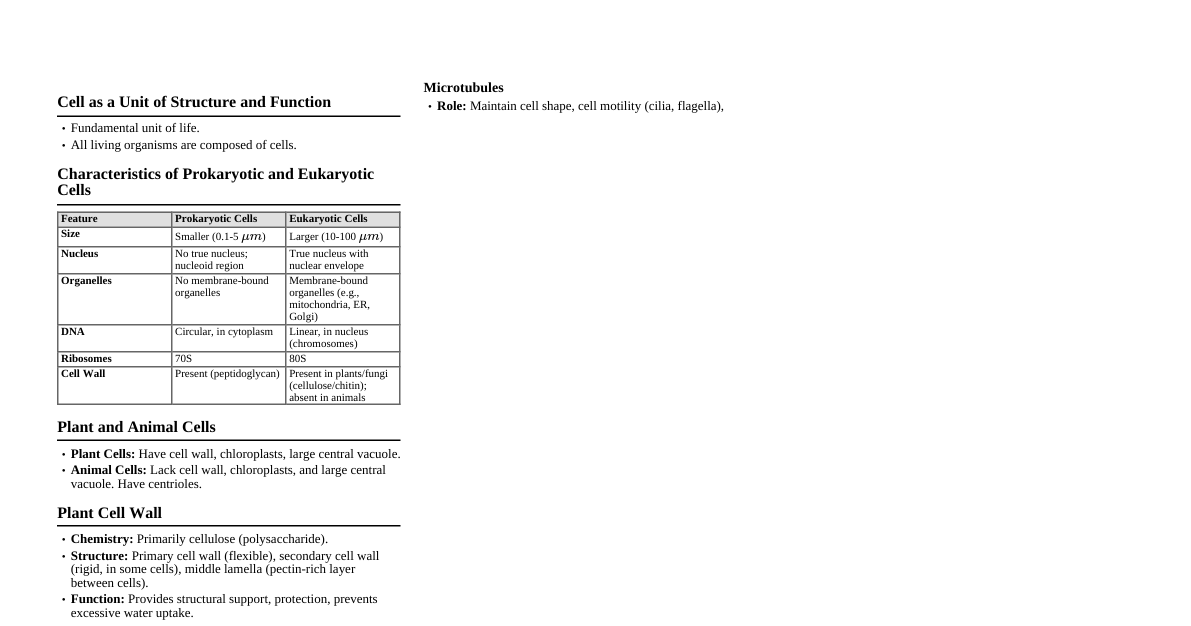
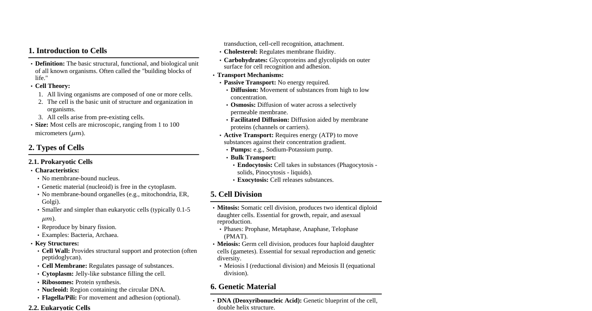
1. Introduction to Cells Definition: The fundamental structural and functional unit of all known living organisms. The smallest unit of life that can replicate independently. Cell Theory: All living organisms are composed of one or more cells. The cell is the basic unit of structure and organization in organisms. All cells arise from pre-existing cells. Two Main Types: Prokaryotic Cells: No true nucleus; genetic material (nucleoid) is in the cytoplasm. Lack membrane-bound organelles. Smaller and simpler (e.g., bacteria, archaea). Possess a cell wall, cell membrane, cytoplasm, ribosomes, and often flagella/pili. Eukaryotic Cells: Possess a true nucleus (enclosed by a nuclear envelope). Contain membrane-bound organelles (e.g., ER, Golgi, mitochondria). Larger and more complex (e.g., animals, plants, fungi, protists). Exhibit compartmentalization, allowing for specialized functions. Key Components Shared by All Cells: Cell membrane, cytoplasm (cytosol + organelles), genetic material (DNA), ribosomes. 2. Cell Membrane (Plasma Membrane) Structure: Fluid Mosaic Model: Describes the membrane as a fluid structure with a "mosaic" of various proteins embedded in or attached to a double layer (bilayer) of phospholipids. Phospholipid Bilayer: Amphipathic molecules with hydrophilic (water-loving) phosphate heads facing the aqueous environment and hydrophobic (water-fearing) fatty acid tails facing each other in the interior. Proteins: Integral Proteins: Span the entire membrane (transmembrane) or are embedded within it. Peripheral Proteins: Loosely bound to the surface of the membrane. Carbohydrates: Often attached to proteins (glycoproteins) or lipids (glycolipids) on the outer surface, forming the glycocalyx (cell recognition). Cholesterol: (Animal cells) Regulates membrane fluidity; reduces fluidity at warmer temperatures and prevents tight packing at colder temperatures. Function: Forms a selectively permeable barrier, controlling what enters and exits the cell. Cell recognition and adhesion (via glycoproteins and glycolipids). Signal transduction (receptor proteins bind to signaling molecules). Enzymatic activity (some membrane proteins act as enzymes). Attachment to the cytoskeleton and extracellular matrix. Transport Mechanisms: Passive Transport: Movement down a concentration gradient; no energy (ATP) required. Diffusion: Movement of substances from high to low concentration. Facilitated Diffusion: Diffusion aided by transport proteins (channel or carrier proteins). Osmosis: Diffusion of water across a selectively permeable membrane. Tonicity: Isotonic, Hypotonic, Hypertonic solutions. Active Transport: Movement against a concentration gradient; requires energy (ATP). Primary Active Transport: Uses ATP directly (e.g., Na+/K+ pump). Secondary Active Transport (Co-transport): Uses energy from an electrochemical gradient established by primary active transport. Bulk Transport: Endocytosis: Cell takes in substances by forming vesicles. Phagocytosis ("cell eating"): Engulfs large particles. Pinocytosis ("cell drinking"): Engulfs extracellular fluid and solutes. Receptor-mediated endocytosis: Specific uptake of molecules using receptors. Exocytosis: Cell releases substances by fusing vesicles with the plasma membrane. 3. Nucleus Structure: Nuclear Envelope: Double membrane (inner and outer) perforated by nuclear pores. Continuous with the ER. Nuclear Pores: Regulate the passage of macromolecules (proteins, RNA) between the nucleus and cytoplasm. Nuclear Lamina: A net-like array of protein filaments (intermediate filaments) lining the inner surface of the nuclear envelope; maintains nuclear shape. Nucleoplasm: The viscous fluid filling the nucleus. Chromatin: Complex of DNA and proteins (histones). DNA is organized into chromosomes during cell division. Nucleolus: A dense, non-membranous structure within the nucleus, responsible for ribosomal RNA (rRNA) synthesis and assembly of ribosomal subunits. Function: Houses and protects the cell's genetic material (DNA). Controls gene expression, cell growth, metabolism, and reproduction. Site of DNA replication and transcription (synthesis of mRNA, tRNA, rRNA). 4. Ribosomes Structure: Small particles composed of ribosomal RNA (rRNA) and ribosomal proteins. Consist of a large subunit and a small subunit. Location: Free Ribosomes: Suspended in the cytosol; synthesize proteins that function within the cytosol (e.g., enzymes, structural proteins). Bound Ribosomes: Attached to the ER or nuclear envelope; synthesize proteins destined for secretion, insertion into membranes, or delivery to certain organelles (e.g., lysosomes, Golgi). Function: Protein synthesis (translation) – translating messenger RNA (mRNA) into polypeptide chains. 5. Endoplasmic Reticulum (ER) Structure: Extensive network of interconnected membranes forming sacs (cisternae) and tubules throughout the cytoplasm. Continuous with the outer nuclear membrane. Types: Rough ER (RER): Appearance: Studded with ribosomes on its cytosolic surface. Function: Synthesis of proteins destined for secretion, insertion into membranes, or delivery to organelles like the Golgi, lysosomes, and vacuoles. Involved in protein folding, modification (e.g., glycosylation), and quality control. Membrane factory for the cell. Smooth ER (SER): Appearance: Lacks ribosomes; appears smooth. Function: Synthesis of lipids (e.g., steroids, phospholipids, oils). Detoxification of drugs, alcohol, and poisons (especially in liver cells). Storage of calcium ions ($Ca^{2+}$) (crucial for muscle contraction and other cellular responses). Metabolism of carbohydrates. 6. Golgi Apparatus (Golgi Complex/Body) Structure: Consists of flattened membrane-bound sacs called cisternae, arranged in stacks. Has distinct structural and functional regions: Cis-face: "Receiving" side, usually located near the ER. Medial-cisternae: Intermediate compartments. Trans-face: "Shipping" side, where materials exit in vesicles. Function: Modifies, sorts, and packages proteins and lipids synthesized in the ER. Adds molecular tags (e.g., phosphate groups) for directing vesicles to specific destinations. Synthesizes certain macromolecules itself (e.g., some polysaccharides in plant cell walls). Produces lysosomes and peroxisomes. Vesicles bud off the trans-face, transporting contents to other organelles or the plasma membrane for secretion. 7. Lysosomes Structure: Spherical membrane-bound sacs containing a variety of hydrolytic (digestive) enzymes. Maintain an acidic internal pH ($\approx 5$) optimal for enzyme activity. Function: Intracellular Digestion: Break down macromolecules (proteins, fats, polysaccharides, nucleic acids) ingested by phagocytosis. Autophagy: Recycle the cell's own damaged organelles and macromolecules. Apoptosis: Involved in programmed cell death (e.g., removal of webbing between fingers and toes during embryonic development). Origin: Formed by budding from the Golgi apparatus. 8. Peroxisomes Structure: Small, membrane-bound organelles containing enzymes, particularly oxidases and catalase. Function: Contain enzymes that remove hydrogen atoms from various substrates and transfer them to oxygen, producing hydrogen peroxide ($H_2O_2$) as a byproduct. Detoxification: Break down fatty acids into smaller molecules for cellular respiration. Detoxify alcohol and other harmful compounds in liver cells. The enzyme catalase converts toxic $H_2O_2$ into water and oxygen ($2H_2O_2 \rightarrow 2H_2O + O_2$). In plant cells, glyoxysomes (a specialized type of peroxisome) convert fatty acids to sugars for germinating seeds. Origin: Grow by incorporating proteins and lipids from the cytosol and ER, and by dividing from pre-existing peroxisomes. 9. Mitochondria Structure: Often referred to as the "powerhouse of the cell." Double Membrane: Outer Membrane: Smooth and permeable to small molecules. Inner Membrane: Highly folded into cristae, increasing surface area for ATP synthesis. Contains electron transport chain components and ATP synthase. Intermembrane Space: The narrow space between the outer and inner membranes. Mitochondrial Matrix: The innermost compartment, containing enzymes for the Krebs cycle, mitochondrial DNA (mtDNA), ribosomes, and various ions. Function: Site of cellular respiration, generating the majority of the cell's ATP (adenosine triphosphate) through oxidative phosphorylation. Involved in calcium signaling, heat production, and apoptosis. Endosymbiont Theory: Mitochondria (and chloroplasts) are thought to have evolved from free-living prokaryotes that were engulfed by ancestral eukaryotic cells. Evidence includes their own circular DNA, ribosomes, and ability to self-replicate. 10. Cytoskeleton Structure: A complex network of protein filaments and tubules extending throughout the cytoplasm of eukaryotic cells. Provides structural support and mechanical strength. Function: Maintains cell shape and provides mechanical support. Facilitates cell motility (movement of the entire cell). Aids in the movement of organelles and vesicles within the cell. Involved in cell division (chromosome segregation, cytokinesis). Components: Microtubules: Structure: Hollow cylinders composed of tubulin protein dimers ($\alpha$-tubulin and $\beta$-tubulin). Have a diameter of about 25 nm. Function: Maintain cell shape (resist compression). Cell motility (as components of cilia and flagella). Chromosome movements in cell division (form the spindle fibers). Organelle movements (act as "tracks" for motor proteins like kinesin and dynein). Associated Structures: Centrosomes, centrioles, basal bodies. Microfilaments (Actin Filaments): Structure: Solid rods composed of actin protein monomers, often arranged in a twisted double chain. Have a diameter of about 7 nm. Function: Maintain cell shape (bear tension). Changes in cell shape (e.g., amoeboid movement). Muscle contraction (interact with myosin). Cytoplasmic streaming in plant cells. Cleavage furrow formation during animal cell division. Projection of cell surface (microvilli). Intermediate Filaments: Structure: Fibrous proteins supercoiled into thicker cables. Diameter of 8-12 nm, intermediate in size. Composed of various proteins (e.g., keratins, lamins), depending on cell type. Function: Maintain cell shape (bear tension). Anchor organelles (e.g., nucleus via nuclear lamina). More permanent and less dynamic than microtubules and microfilaments. Form the nuclear lamina. 11. Centrosomes and Centrioles (Animal Cells and some lower plants) Structure: Centrosome: A region located near the nucleus, considered the main microtubule-organizing center (MTOC) in animal cells. Centrioles: Two centrioles are found within the centrosome, arranged perpendicularly to each other. Each centriole is composed of nine triplets of microtubules arranged in a ring. Function: Organize microtubules within the cell. Involved in cell division: replicate during interphase, then move to opposite poles of the cell to organize the mitotic spindle. Form the basal bodies of cilia and flagella. 12. Cell Wall (Plant, Fungi, Bacteria, Algae) Structure: A rigid layer outside the plasma membrane. Plants: Primarily composed of cellulose microfibrils embedded in a matrix of other polysaccharides and proteins. Primary cell wall (thin, flexible) and secondary cell wall (thicker, stronger). Fungi: Made of chitin. Bacteria: Made of peptidoglycan. Algae: Variable composition (e.g., cellulose, agar, carrageenan). Function: Provides structural support and protection to the cell. Maintains cell shape. Prevents excessive water uptake (osmotic lysis) by exerting turgor pressure against the plasma membrane. Acts as a barrier against pathogens. 13. Vacuoles Structure: Large, membrane-bound sacs within the cytoplasm. The membrane is called the tonoplast. Types and Functions: Central Vacuole (Plant Cells): Typically large and occupies 30-80% or more of the cell volume. Stores water, nutrients, ions, pigments, and waste products. Maintains turgor pressure against the cell wall, supporting the plant. Can act as a lysosome for intracellular digestion. Food Vacuoles: Formed by phagocytosis; fuse with lysosomes for digestion. Contractile Vacuoles (Protists): Pump excess water out of the cell, maintaining water balance (osmoregulation). Storage Vacuoles: Store various substances (e.g., seed storage proteins in plant seeds). 14. Chloroplasts (Plant and Algal Cells) Structure: Member of the plastid family. Double Membrane: Outer and inner membranes. Stroma: The fluid-filled space inside the inner membrane, containing enzymes for the Calvin cycle, chloroplast DNA (cpDNA), and ribosomes. Thylakoids: Flattened, interconnected sacs within the stroma. The site of the light-dependent reactions of photosynthesis. Grana (singular: granum): Stacks of thylakoids. Chlorophyll: The green pigment located in the thylakoid membranes, absorbs light energy. Function: Site of photosynthesis – converts light energy into chemical energy (sugars) using carbon dioxide and water. $6CO_2 + 6H_2O + \text{Light energy} \rightarrow C_6H_{12}O_6 + 6O_2$ Endosymbiont Theory: Like mitochondria, chloroplasts are believed to have originated from an endosymbiotic relationship with a photosynthetic prokaryote. 15. Intercellular Junctions Function: Connect cells to each other and to the extracellular matrix, facilitating communication and tissue integrity. Types (Animal Cells): Tight Junctions (Occluding Junctions): Structure: Formed by proteins (e.g., claudins, occludins) that press adjacent plasma membranes together. Function: Create a watertight seal, preventing the leakage of extracellular fluid across a layer of epithelial cells (e.g., in the gut lining, blood-brain barrier). Desmosomes (Anchoring Junctions): Structure: Protein filaments (intermediate filaments, specifically keratin) extend into the cytoplasm, anchoring desmosomes in adjacent cells. Function: Fasten cells together into strong sheets, providing mechanical strength (e.g., in muscle tissue, skin). Resist pulling forces. Gap Junctions (Communicating Junctions): Structure: Consist of protein channels (connexons) that span the plasma membranes of adjacent cells, creating direct cytoplasmic connections. Function: Allow for rapid passage of small molecules (ions, sugars, amino acids) and electrical signals between cells. Crucial for coordinated activity (e.g., heart muscle, embryonic development). Types (Plant Cells): Plasmodesmata (Singular: plasmodesma): Structure: Channels that perforate plant cell walls, connecting the cytoplasm of adjacent plant cells. The plasma membrane is continuous through these channels. Function: Allow for the passage of water, small solutes, and some larger molecules (e.g., proteins, RNA) between cells, enabling intercellular communication and transport.