Intestinal Diseases (K. Park 28th Ed)
Cheatsheet Content
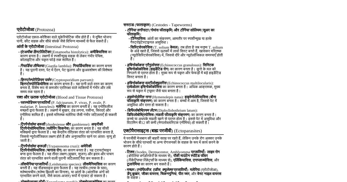
1. Diarrheal Diseases Definition: Passage of 3 or more loose or liquid stools per day, or more frequently than normal for the individual. Diarrhea is a major public health problem, especially in developing countries, leading cause of morbidity and mortality in children under 5. Types: Acute watery diarrhea: Lasts several hours or days, includes cholera. Leads to dehydration. Acute bloody diarrhea (dysentery): Lasts several hours or days, associated with fever and abdominal pain. Suggests invasive pathogens. Persistent diarrhea: Lasts 14 days or longer. Often associated with malnutrition and malabsorption. Chronic diarrhea: Lasts >4 weeks. Often non-infectious causes but can be parasitic. Etiology: Bacterial: Toxigenic: Vibrio cholerae (Cholera toxin - CT), Enterotoxigenic E. coli (ETEC - LT/ST toxins), Clostridium perfringens , Staphylococcus aureus , Bacillus cereus . Primarily cause watery diarrhea. Invasive/Cytotoxic: Shigella spp. (Shiga toxin), Enteroinvasive E. coli (EIEC), Salmonella spp. (non-typhoidal), Campylobacter jejuni , Enterohemorrhagic E. coli (EHEC/STEC - Shiga-like toxin, e.g., O157:H7). Primarily cause dysentery/bloody diarrhea. Viral: Rotavirus (most common cause of severe diarrhea in infants/young children), Norovirus (common in outbreaks, all ages), Adenovirus, Astrovirus. Cause watery diarrhea. Parasitic: Giardia lamblia , Entamoeba histolytica , Cryptosporidium parvum , Cyclospora cayetanensis . Can cause acute, persistent, or chronic diarrhea. Risk Factors: Contaminated food/water, poor personal and community hygiene, lack of sanitation, malnutrition, immunodeficiency (HIV/AIDS), crowded living conditions. Pathophysiology: Secretory: Toxins stimulate active secretion of water and electrolytes into the lumen (e.g., cholera, ETEC). Osmotic: Non-absorbable solutes in the lumen draw water (e.g., lactose intolerance, malabsorption). Inflammatory/Invasive: Damage to mucosal cells, ulceration, exudation (e.g., Shigella, EIEC, EHEC, Entamoeba). Malabsorption: Damage to villi, reduced surface area (e.g., Giardia, persistent diarrhea). Prevention: Intervention at Source: Safe Water: Chlorination, boiling, filtration, protected water sources. Safe Food: Proper food handling, cooking, storage (e.g., "5 Keys to Safer Food" - WHO). Sanitation: Safe disposal of human and animal feces, construction and use of latrines. Disease Surveillance: Early detection of outbreaks. Intervention at Mode of Transmission: Handwashing: With soap and water, especially after defecation and before handling food/eating. Vector Control: Flies (physical barriers, insecticides). Personal Hygiene: Bathing, clean clothes. Avoidance of Contaminated Sources: Don't eat raw/undercooked food, avoid untreated water. Intervention at Host: Vaccination: Rotavirus vaccine for infants, Oral Cholera Vaccine (OCV), Typhoid vaccines. Exclusive Breastfeeding: For first 6 months of life. Improved Nutrition: Reduces susceptibility and severity. Health Education: Promoting hygienic practices. Early Diagnosis & Treatment: Prompt ORS and appropriate antibiotics/anthelmintics. Nutritional Rehabilitation: For persistent diarrhea to prevent malnutrition. Treatment (WHO Integrated Management of Childhood Illness - IMCI): Plan A: Treat Diarrhea at Home (No Dehydration) Assessment: No signs of dehydration (no thirst, normal skin pinch, normal eyes/tears, moist mouth). Key Actions: Give more fluid than usual: Encourage oral fluids like ORS (if available), homemade solutions (salt-sugar solution), gruel, rice water, yogurt drink, soup, fresh fruit juice. Avoid sugary drinks. Continue feeding: Breastfeed frequently. For non-breastfed children, continue regular diet. Give frequent, small meals (6 times/day). Zinc supplementation: $10 \text{ mg/day}$ for children $ Teach mother when to return immediately: Danger signs include passing many watery stools, repeated vomiting, drinking poorly or not at all, getting sicker, fever, blood in stool. Plan B: Treat Dehydration with ORS (Some Dehydration) Assessment: Two or more signs of dehydration (restless/irritable, sunken eyes, drinks eagerly (thirsty), skin pinch goes back slowly). Key Actions: Administer ORS solution orally in a health facility (or at home if instructed). Use WHO low-osmolarity ORS ($245 \text{ mOsm/L}$). Amount of ORS to give over 4 hours: Up to 4 months ($ 4 to 12 months ($5-7.9 \text{ kg}$): $400-600 \text{ ml}$ 12 to 24 months ($8-10.9 \text{ kg}$): $600-800 \text{ ml}$ 2 to 5 years ($11-15.9 \text{ kg}$): $800-1200 \text{ ml}$ $\geq 5$ years ($\geq 16 \text{ kg}$): $1200-2200 \text{ ml}$ Adults: $2-4 \text{ L}$ over 4 hours. Reassess after 4 hours: If no dehydration, switch to Plan A. If some dehydration persists, repeat Plan B. If severe dehydration develops, proceed to Plan C. Continue breastfeeding during ORS administration. Zinc supplementation as above. Plan C: Treat Severe Dehydration Quickly (Severe Dehydration) Assessment: Two or more signs of severe dehydration (lethargic/unconscious, sunken eyes, drinks poorly or not at all, skin pinch goes back very slowly (>2 seconds)). Key Actions: Administer IV fluids immediately: Ringer's Lactate (Hartmann's solution) is preferred. If not available, Normal Saline. Rapid infusion schedule: Infants ($ $30 \text{ ml/kg}$ in 1 hour, then $70 \text{ ml/kg}$ in 5 hours. Children ($\geq 12$ months) & Adults: $30 \text{ ml/kg}$ in 30 minutes, then $70 \text{ ml/kg}$ in 2.5 hours. Reassess frequently: Every 15-30 minutes for infants, every 30-60 minutes for older children/adults. Check pulse, consciousness, skin pinch. As soon as patient can drink: Give ORS ($5 \text{ ml/kg/hr}$) along with IV fluids. After 6 hours (infants) or 3 hours (older children/adults): Reassess and switch to Plan A or B as appropriate. Zinc supplementation as above. Antibiotics: For specific cases like cholera (doxycycline) or severe dysentery (ceftriaxone). Antibiotics for Diarrhea: Generally NOT recommended for acute watery diarrhea (unless cholera suspected) due to viral etiology, risk of resistance, and adverse effects. Dysentery (bloody diarrhea): Empiric treatment with Ciprofloxacin (adults) or Ceftriaxone (children) is often initiated, pending stool culture. Azithromycin is an alternative for resistant Shigella. Cholera: Doxycycline, Azithromycin, Ciprofloxacin (shortens duration and reduces infectivity). Persistent Diarrhea: May require investigation for parasitic causes (Giardia, Cryptosporidium) or bacterial overgrowth. 2. Cholera Agent: Vibrio cholerae serogroups O1 (classic and El Tor biotypes) and O139. Gram-negative, comma-shaped bacillus. Pathogenesis: Cholera toxin (CT) is an A-B toxin that activates adenylate cyclase in intestinal epithelial cells, leading to increased cAMP and massive secretion of fluid and electrolytes. Transmission: Feco-oral route, primarily through contaminated water (epidemic) or food (sporadic cases). Incubation period: few hours to 5 days, typically 2-3 days. Clinical Features: Sudden onset of profuse, painless, watery diarrhea ("rice-water stools" - flecks of mucus, no blood). Rapid progression to severe dehydration, hypovolemic shock. Vomiting (non-bilious) often follows diarrhea. Muscle cramps (due to electrolyte loss), decreased urine output (anuria), sunken eyes, dry mucous membranes, loss of skin turgor. Pulse may be weak/absent, blood pressure unrecordable. Severe cases can lead to death within hours if untreated. Diagnosis: Clinical suspicion: In endemic areas or during outbreaks. Stool microscopy: Darting motility of vibrios. Culture: Thiosulphate Citrate Bile-salt Sucrose (TCBS) agar (yellow colonies). Definitive. Rapid diagnostic tests (RDTs): Dipsticks for O1/O139 antigens (useful for surveillance). Treatment: Rapid rehydration is paramount: Severe dehydration: IV fluids (Ringer's Lactate) per Plan C. Mild/moderate dehydration: ORS per Plan B. Antibiotics: Shorten duration of diarrhea, reduce fluid loss, and decrease the period of vibrio excretion. Adults: Doxycycline $300 \text{ mg}$ single oral dose. Alternative: Ciprofloxacin $1 \text{ g}$ single oral dose. Children: Azithromycin $20 \text{ mg/kg}$ single oral dose. Alternative: Erythromycin. Resistance patterns vary geographically. Zinc supplementation: For children. Prevention: Source: Safe water supply, proper sanitation (safe disposal of human feces). Mode of Transmission: Food hygiene (cooking food thoroughly, avoiding raw foods in endemic areas), handwashing. Host: Oral Cholera Vaccines (OCV) for targeted populations or travelers, health education. 3. Typhoid Fever (Enteric Fever) Agent: Salmonella Typhi (most common) and Salmonella Paratyphi A, B, C. Gram-negative bacillus. Pathogenesis: Ingestion $\rightarrow$ invade Peyer's patches $\rightarrow$ regional lymph nodes $\rightarrow$ bloodstream (primary bacteremia) $\rightarrow$ reticuloendothelial system (liver, spleen, bone marrow) $\rightarrow$ secondary bacteremia $\rightarrow$ recolonization of Peyer's patches and gallbladder (carrier state). Transmission: Feco-oral route, contaminated food/water, chronic carriers (e.g., "Typhoid Mary"). Incubation period: 7-14 days. Clinical Features (Classic presentation, may be atypical): Week 1: Gradual onset of "step-ladder" fever (rises daily), headache, malaise, anorexia, relative bradycardia, constipation (more common than diarrhea initially). Week 2: Sustained high fever, apathy, "rose spots" (faint, salmon-colored maculopapular rash on trunk/abdomen, blanch with pressure), hepatosplenomegaly, "pea-soup" diarrhea, abdominal tenderness. Week 3: If untreated, complications arise. Worsening toxemia, delirium. Complications: Intestinal hemorrhage (most common, due to necrosis of Peyer's patches), intestinal perforation (most serious, leads to peritonitis), encephalopathy, myocarditis, cholecystitis, osteomyelitis, meningitis. Chronic carrier state: Excretion of bacteria for >1 year, usually from gallbladder. Diagnosis: Blood culture: Gold standard. Positive in 70-80% in first week. Bone marrow culture: Highest sensitivity (90%), useful in partially treated cases. Stool culture: Positive later, useful for identifying carriers. Urine culture: Less sensitive. Widal test: Detects agglutinating antibodies (O and H antigens). Retrospective value, poor sensitivity/specificity due to prior infection, vaccination, or cross-reactivity. A four-fold rise in titer in paired sera is more significant. Typhidot/Typhidot M: Detects IgM/IgG antibodies to S. Typhi (IgM suggests acute infection). Rapid test. Treatment: Antibiotics (key to treatment): Duration 7-14 days, depending on severity and agent. Drug-Sensitive Strains: Ciprofloxacin $500 \text{ mg}$ BID OR Azithromycin $500 \text{ mg}$ once daily. Multidrug-Resistant (MDR) Strains (resistant to chloramphenicol, ampicillin, cotrimoxazole): Fluoroquinolones (Ciprofloxacin, Ofloxacin) if susceptible. Third-generation Cephalosporins: Ceftriaxone $50-75 \text{ mg/kg/day}$ IV/IM for 10-14 days. Azithromycin $1 \text{ g}$ on day 1 then $500 \text{ mg/day}$ for 6 days (or $20 \text{ mg/kg/day}$ for 7 days in children). Good oral bioavailability. Extensively Drug-Resistant (XDR) Strains (resistant to FQ and 3rd gen ceph): Azithromycin, Carbapenems (Meropenem). Old drugs (if susceptible and no resistance): Chloramphenicol, Ampicillin, Cotrimoxazole. Supportive care: Fluid and electrolyte balance, nutrition, antipyretics. Steroids: Dexamethasone for severe toxemia, encephalopathy, shock. Surgery: For intestinal perforation. Prevention: Source: Safe water, proper sanitation (safe disposal of human feces), identification and treatment of chronic carriers. Mode of Transmission: Food hygiene, handwashing, avoiding raw foods. Host: Typhoid vaccines (Vi polysaccharide, Ty21a oral, TCV), health education. 4. Helminthic Infections (Intestinal Worms) General Features: Soil-transmitted helminths (STHs) are transmitted through contaminated soil. High prevalence in areas with poor sanitation. Cause chronic infections, often lead to malnutrition, anemia, impaired cognitive development. Ascariasis (Roundworm): Agent: Ascaris lumbricoides . Largest intestinal nematode. Life Cycle: Ingestion of embryonated eggs $\rightarrow$ larvae hatch in small intestine $\rightarrow$ penetrate intestinal wall $\rightarrow$ portal circulation $\rightarrow$ liver $\rightarrow$ heart $\rightarrow$ lungs (molt) $\rightarrow$ ascend bronchi/trachea $\rightarrow$ swallowed $\rightarrow$ mature in small intestine, lay eggs. Clinical: Asymptomatic in light infections. Lung phase: Ascaris pneumonitis (Loeffler's syndrome): cough, dyspnea, eosinophilia. Intestinal phase: Abdominal pain, malnutrition, malabsorption. Heavy infections can cause intestinal obstruction, volvulus. Migration of adult worms: Biliary obstruction, pancreatitis, appendicitis, expulsion through mouth/nose. Diagnosis: Stool microscopy for characteristic mammillated eggs. Adult worms in stool/vomitus. Treatment: Albendazole $400 \text{ mg}$ single dose OR Mebendazole $500 \text{ mg}$ single dose OR $100 \text{ mg}$ BID for 3 days. Pyrantel Pamoate (safe in pregnancy). Prevention: Source: Safe disposal of human feces. Mode of Transmission: Food hygiene, handwashing, avoiding contaminated soil. Host: Mass drug administration (MDA) in endemic areas, health education. Hookworm (Ancylostomiasis/Necatoriasis): Agents: Ancylostoma duodenale , Necator americanus . Life Cycle: Filariform larvae in soil penetrate skin (usually feet) $\rightarrow$ bloodstream $\rightarrow$ lungs $\rightarrow$ ascend trachea $\rightarrow$ swallowed $\rightarrow$ mature in small intestine, attach to mucosa, feed on blood. Clinical: Skin: "Ground itch" (pruritic papules/vesicles) at larval entry site. Lung: Mild cough, wheezing during larval migration. Intestinal: Iron deficiency anemia (due to chronic blood loss), abdominal pain, protein-losing enteropathy, growth retardation. Diagnosis: Stool microscopy for characteristic oval eggs. Treatment: Albendazole $400 \text{ mg}$ single dose OR Mebendazole $500 \text{ mg}$ single dose OR $100 \text{ mg}$ BID for 3 days. Iron supplementation for anemia. Prevention: Source: Safe disposal of human feces. Mode of Transmission: Wearing shoes, avoiding contact with contaminated soil. Host: MDA in endemic areas, health education. Trichuriasis (Whipworm): Agent: Trichuris trichiura . Life Cycle: Ingestion of embryonated eggs $\rightarrow$ larvae hatch in small intestine $\rightarrow$ mature in large intestine (cecum/colon), embed anterior portion in mucosa. Clinical: Often asymptomatic in light infections. Heavy infections: Abdominal pain, bloody diarrhea, tenesmus, weight loss, malnutrition, growth retardation. Rectal prolapse (classic in children with heavy infection). Diagnosis: Stool microscopy for characteristic barrel-shaped eggs with polar plugs. Treatment: Mebendazole $100 \text{ mg}$ BID for 3 days OR Albendazole $400 \text{ mg}$ daily for 3 days. Prevention: Source: Safe disposal of human feces. Mode of Transmission: Food hygiene, handwashing, avoiding contaminated soil. Host: MDA in endemic areas, health education. Enterobiasis (Pinworm/Threadworm): Agent: Enterobius vermicularis . Life Cycle: Ingestion of eggs $\rightarrow$ larvae hatch in small intestine $\rightarrow$ mature in large intestine $\rightarrow$ gravid females migrate to perianal region at night to lay eggs. Clinical: Perianal itching (pruritus ani) , especially nocturnal. Insomnia, irritability, secondary bacterial infection due to scratching. Rarely, vulvovaginitis or ectopic infections. Diagnosis: Scotch tape test (perianal swab) in the morning before bathing/defecation. Eggs are rarely seen in stool. Treatment: Mebendazole $100 \text{ mg}$ single dose OR Albendazole $400 \text{ mg}$ single dose. Repeat dose after 2 weeks to kill newly hatched larvae. Treat entire household. Prevention: Source: Human feces (eggs). Mode of Transmission: Handwashing (especially after using toilet and before eating), regular washing of bed linens/undergarments, good personal hygiene. Host: Treatment of entire household, health education. Taeniasis (Tapeworm): Agents: Taenia saginata (beef tapeworm), Taenia solium (pork tapeworm). Life Cycle (Taeniasis): Ingestion of raw/undercooked meat containing cysticerci (larval cysts) $\rightarrow$ adult worm develops in small intestine. Clinical (Taeniasis): Often asymptomatic, mild abdominal discomfort, nausea, passage of motile proglottids in stool (especially T. saginata ). Diagnosis: Stool examination for proglottids or eggs. Eggs are morphologically identical for T. saginata and T. solium . Treatment (Taeniasis): Praziquantel $5-10 \text{ mg/kg}$ single dose. Niclosamide (alternative). Cysticercosis (specific to T. solium ): Life Cycle (Cysticercosis): Ingestion of T. solium eggs (from human feces) $\rightarrow$ oncospheres hatch $\rightarrow$ disseminate to tissues (muscle, brain, eye) $\rightarrow$ form cysticerci. Humans are accidental intermediate host. Neurocysticercosis: Most severe form. Seizures, headache, hydrocephalus, focal neurological deficits. Diagnosis of Cysticercosis: Imaging (CT/MRI for neurocysticercosis), serology (ELISA for antibodies). Treatment of Cysticercosis: Albendazole + Praziquantel (for viable cysts), steroids (to reduce inflammation from dying cysts), anti-epileptics for seizures, surgery for hydrocephalus/large cysts. Prevention: Source: Infected cattle/pigs (for taeniasis), human feces (for cysticercosis). Mode of Transmission: Proper cooking of meat (taeniasis), handwashing, safe disposal of human feces (cysticercosis). Host: Meat inspection, health education. 5. Giardiasis Agent: Giardia lamblia (also known as G. intestinalis or G. duodenalis ). A flagellated protozoan. Life Cycle: Ingestion of cysts (infective stage) $\rightarrow$ trophozoites emerge in small intestine $\rightarrow$ attach to intestinal villi (do not invade) $\rightarrow$ multiply by binary fission $\rightarrow$ encyst in colon $\rightarrow$ cysts passed in feces. Transmission: Feco-oral route, contaminated water ("beaver fever"), food, person-to-person. Low infectious dose. Clinical Features: Acute: Diarrhea (often greasy, foul-smelling, non-bloody), abdominal cramps, bloating, flatulence, nausea, malaise, weight loss. Chronic: Intermittent diarrhea, malabsorption (fat, fat-soluble vitamins, lactose), weight loss, growth faltering in children. Can mimic irritable bowel syndrome. Often asymptomatic carriage. Pathology: Villous atrophy, crypt hyperplasia, inflammatory infiltration of lamina propria, reduced brush border enzymes. Diagnosis: Stool examination: Microscopic identification of cysts (oval) or trophozoites (pear-shaped with flagella) in fresh or concentrated stool. Multiple samples (3 over several days) increase yield. Antigen detection: ELISA or rapid immunochromatographic tests on stool (high sensitivity/specificity). Direct fluorescent antibody (DFA) for cysts/trophozoites. Entero-test (string test) / Duodenal aspirate: Rarely used, but can detect trophozoites. Treatment: Metronidazole $250 \text{ mg}$ TID for 5-7 days (adults), $15 \text{ mg/kg/day}$ in 3 doses for 5-7 days (children). Tinidazole $2 \text{ g}$ single dose (adults), $50 \text{ mg/kg}$ single dose (children) - often preferred due to single dose. Nitazoxanide (broad-spectrum, for children). Albendazole (less efficacious than metronidazole, but alternative). Prevention: Source: Contaminated water sources (ponds, streams), human/animal feces. Mode of Transmission: Boiling/filtering drinking water, handwashing, proper food preparation. Host: Health education. 6. Amebiasis Agent: Entamoeba histolytica . Pathogenic amoeba. Note: E. dispar is morphologically identical but non-pathogenic. Life Cycle: Ingestion of cysts (infective stage) $\rightarrow$ trophozoites emerge in small intestine $\rightarrow$ colonize large intestine. Trophozoites invade colonic mucosa causing disease, or encyst and are passed in feces. Transmission: Feco-oral route, contaminated food/water, person-to-person, flies, sexual contact (MSM). Clinical Features: Asymptomatic carriage: Most common (90%), often due to non-pathogenic E. dispar or resistant host. Intestinal Amoebiasis: Amoebic Colitis/Dysentery: Gradual onset of bloody, mucoid diarrhea ("raspberry jelly stool"), abdominal pain (colicky), tenesmus, fever (less common than bacterial dysentery). Flask-shaped ulcers in colon. Non-dysenteric colitis: Milder diarrhea, abdominal pain. Ameboma: Granulomatous mass in colon, can mimic carcinoma. Fulminant amoebic colitis: Severe, toxic megacolon, perforation. Extra-intestinal Amoebiasis: Amoebic Liver Abscess (ALA): Most common extra-intestinal manifestation. Right upper quadrant pain, fever, hepatomegaly, weight loss. "Anchovy paste" pus. More common in males. Less common sites: Lung (pleuropulmonary), brain, skin (perianal). Diagnosis: Stool examination: Microscopic identification of trophozoites (with ingested RBCs, motile) in fresh dysenteric stool OR cysts (quadrinucleate) in formed stool. Differentiation from E. dispar is crucial (use PCR/antigen tests). Antigen detection: ELISA for E. histolytica antigen in stool (differentiates from E. dispar ). Serology (Indirect Hemagglutination - IHA, ELISA): Useful for invasive disease (ALA), as antibodies are often absent in asymptomatic carriers. Imaging for ALA: Ultrasound, CT scan (single, round/oval lesion, typically in right lobe). Aspiration of liver abscess: "Anchovy paste" pus, sterile on bacterial culture. Treatment: For Invasive Amoebiasis (amoebic colitis, liver abscess): Tissue amoebicides: Metronidazole $750 \text{ mg}$ TID for 5-10 days (adults), $35-50 \text{ mg/kg/day}$ in 3 doses for 5-10 days (children). OR Tinidazole. Followed by a luminal amoebicide: To eradicate cysts and prevent relapse/transmission from the colon. For Asymptomatic cyst passers/Luminal treatment (to prevent invasive disease and transmission): Diloxanide furoate $500 \text{ mg}$ TID for 10 days (adults). Paromomycin $25-35 \text{ mg/kg/day}$ in 3 doses for 7 days. Iodoquinol. Aspiration of liver abscess: Generally not needed unless large, impending rupture, or no response to medical therapy. Prevention: Source: Human feces. Mode of Transmission: Safe water, proper sanitation, food hygiene, handwashing, avoiding uncooked food in endemic areas. Host: Health education. 7. Food Poisoning (Foodborne Illness) Definition: Illness caused by consuming food or water contaminated with bacteria, viruses, parasites, or their toxins. Classification: Intoxication: Ingestion of preformed toxins in food. Rapid onset (hours). E.g., S. aureus , B. cereus (emetic toxin). Infection: Ingestion of live organisms which multiply in the gut and cause illness. Longer incubation (days). E.g., Salmonella , Campylobacter , Norovirus. Toxicoinfection: Ingestion of organisms that produce toxins in the gut. E.g., C. perfringens , B. cereus (diarrheal toxin), EHEC. Common Agents & Features: Staphylococcus aureus : Source: Contaminated food by human handlers (salads, pastries, meats). Toxin: Heat-stable enterotoxins. Onset: Very rapid (1-6 hours). Symptoms: Prominent nausea, violent vomiting, abdominal cramps, diarrhea. Fever usually absent. Clostridium perfringens : Source: Cooked meat/poultry dishes left at room temperature. Toxin: Enterotoxin produced in gut. Onset: 8-12 hours. Symptoms: Abdominal cramps, watery diarrhea. Vomiting rare. Bacillus cereus : Source: Emetic type: Reheated rice. Toxin: Cereulide (heat-stable). Onset: 1-5 hours. Symptoms: Nausea, vomiting. Diarrheal type: Meats, vegetables. Toxin: Labile enterotoxin. Onset: 8-16 hours. Symptoms: Diarrhea, abdominal pain. Salmonella spp. (non-typhoidal): Source: Raw/undercooked eggs, poultry, meat, dairy. Onset: 12-72 hours. Symptoms: Nausea, vomiting, abdominal cramps, fever, watery to bloody diarrhea. Campylobacter jejuni : Source: Raw/undercooked poultry, unpasteurized milk, contaminated water. Onset: 2-5 days. Symptoms: Fever, abdominal pain, bloody diarrhea. Can cause Guillain-Barré syndrome post-infection. EHEC (Shiga toxin-producing E. coli , e.g., O157:H7): Source: Undercooked beef, raw milk, contaminated produce. Onset: 3-4 days. Symptoms: Severe abdominal cramps, bloody diarrhea (hemorrhagic colitis), little or no fever. Can lead to Hemolytic Uremic Syndrome (HUS) in children. Norovirus: Source: Shellfish, contaminated water, ready-to-eat foods, person-to-person. Highly contagious. Onset: 12-48 hours. Symptoms: Vomiting (prominent), watery diarrhea, abdominal cramps, low-grade fever. Management: Supportive care: Primarily rehydration with ORS. IV fluids for severe dehydration. Antiemetics: For severe vomiting. Antibiotics: Generally NOT recommended for most food poisoning cases due to viral etiology or toxin-mediated illness. May worsen EHEC by increasing toxin release and HUS risk. Used only in specific bacterial infections (e.g., severe salmonellosis in immunocompromised, traveler's diarrhea). Antidiarrheals: Avoid in bloody diarrhea or suspected invasive infections. Prevention: Source: Contaminated raw foods (meat, poultry, eggs, dairy, produce), infected food handlers. Mode of Transmission: Proper food handling, cooking thoroughly, keeping food at safe temperatures (hot/cold), avoiding cross-contamination, handwashing. Host: Health education on safe food practices.