Cell Division Cheatsheet
Cheatsheet Content
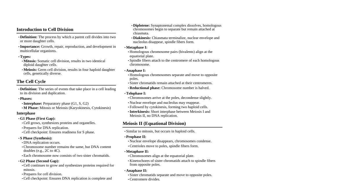
Cell Cycle Overview Sequence of events by which a cell duplicates its genome, synthesizes other cell constituents, and eventually divides into two daughter cells. Human cells: $\approx 24$ hours. Yeast cells: $\approx 90$ minutes. Two main phases: Interphase: Resting phase, cell prepares for division. Accounts for $>95\%$ of the cell cycle duration. M Phase: Actual cell division (mitosis or meiosis). Interphase Cell prepares for division through growth and DNA replication. Divided into three sub-phases: G1 Phase (Gap 1): Interval between mitosis and initiation of DNA replication. Metabolically active, grows continuously. Cell organelle duplication (except DNA and centrioles). Cell decides whether to divide, differentiate, or enter G0. S Phase (Synthesis): DNA replication occurs in the nucleus. Amount of DNA doubles (from $2C$ to $4C$), but chromosome number remains the same (e.g., if $2n$ chromosomes, still $2n$ chromosomes, but each with two chromatids instead of one). Centriole duplication in the cytoplasm. Histone proteins are synthesized. G2 Phase (Gap 2): Proteins necessary for mitosis (e.g., tubulin for spindle fibers) are synthesized. Cell growth continues, cell prepares for division. Checks for DNA damage and ensures DNA replication is complete. G0 Phase (Quiescent Stage) Cells that do not divide further exit G1 phase and enter G0. Metabolically active but no longer proliferate unless signaled to do so. Examples: Heart cells, nerve cells, mature red blood cells (terminally differentiated). Liver cells can re-enter the cell cycle. Cell Cycle Checkpoints Critical control points where "stop" and "go-ahead" signals regulate the cycle. Main checkpoints: G1 Checkpoint (Restriction Point): Most important. Checks for cell size, nutrients, growth factors, DNA damage. If cell passes, it commits to division. G2 Checkpoint: Checks for DNA replication completion and DNA damage. Ensures all chromosomes are replicated and undamaged. M Checkpoint (Spindle Checkpoint): Occurs during metaphase. Ensures all sister chromatids are correctly attached to spindle microtubules before anaphase. Regulated by cyclins and cyclin-dependent kinases (Cdks). Mitosis (M Phase) Dramatic period of cell cycle involving major reorganization of cell components. Equational Division: Number of chromosomes in parent and progeny cells remains the same. Occurs in diploid somatic cells in animals. Occurs in both haploid and diploid cells in plants. Divided into four stages of nuclear division (karyokinesis), followed by cytokinesis. Prophase (Mitosis) Initiation of condensation of chromosomal material, becoming visible under light microscope. Centrosomes (duplicated during S phase) move towards opposite poles, radiating astral rays (microtubules). Chromosomal material condenses to form compact mitotic chromosomes, each composed of 2 sister chromatids attached at the centromere. Mitotic apparatus (spindle fibers, asters) begins to form. Nuclear envelope, nucleolus, Golgi complex, ER disappear ("Prophase to Prometaphase transition"). Metaphase (Mitosis) Complete disintegration of the nuclear envelope. Chromosomes are arranged at the metaphase plate (equator), equidistant from two spindle poles. Morphology of chromosomes (size, shape) is best observed during this stage. Spindle fibers (microtubules) attach to kinetochores (disc-shaped protein structures on the surface of the centromere). Each chromatid has its own kinetochore. Anaphase (Mitosis) Centromere splits (centromeric division), and sister chromatids separate. Sister chromatids (now referred to as daughter chromosomes) move rapidly towards opposite poles due to shortening of kinetochore microtubules. Chromosomes take on V, L, J, or I shapes depending on the position of the centromere (metacentric, submetacentric, acrocentric, telocentric). Telophase (Mitosis) Daughter chromosomes cluster at opposite poles and begin to decondense, losing their individuality. Nuclear envelope develops around each set of chromosomes. Nucleolus, Golgi complex, ER reform inside each newly forming daughter nucleus. The two sets of chromosomes are genetically identical. Cytokinesis (Cytoplasm Separation) The process of cytoplasmic division following nuclear division. Animal Cell: A furrow appears in the plasma membrane (cleavage furrow) due to the contraction of a ring of actin and myosin filaments. This furrow deepens centripetally and eventually divides the cytoplasm. Plant Cell: A new cell wall is formed. Vesicles from the Golgi apparatus fuse at the equator to form a cell plate, which grows centrifugally (outward) until it fuses with the existing lateral walls. The cell plate represents the middle lamella between the two new daughter cells. Syncytium: Karyokinesis not followed by cytokinesis, leading to a multinucleate condition (e.g., liquid endosperm in coconut, skeletal muscle cells). Significance of Mitosis Growth: Responsible for the growth of multicellular organisms from a single zygote. Cell Repair: Replaces dead or damaged cells (e.g., epidermis, lining of the gut, blood cells). Asexual Reproduction: In some lower organisms (e.g., amoeba, yeast), mitosis is the primary mode of reproduction. Maintenance of Nucleo-cytoplasmic Ratio: As a cell grows, its volume increases faster than its surface area, disrupting the ratio. Mitosis restores this ratio. Meiosis Special kind of cell division that reduces chromosome number by half. Ensures production of haploid phase in life cycle of sexually reproducing organisms (e.g., gametes in animals, spores in plants). Involves two sequential cycles of nuclear and cell division (Meiosis I and Meiosis II) but only a single cycle of DNA replication (before Meiosis I). Initiated after parent chromosomes have replicated in S-phase, resulting in each chromosome having two sister chromatids. Involves pairing of homologous chromosomes (synapsis) and genetic recombination (crossing over). Four haploid cells are formed at the end of Meiosis II, which are genetically distinct from the parent cell and from each other. Occurs in meiocytes (germline cells). Meiosis I (Reductional Division) Homologous chromosomes separate, reducing the chromosome number by half. Prophase I Longer and more complex than mitotic prophase. Divided into 5 sub-stages: Leptotene: Chromosomes become gradually visible and start to condense. Each chromosome consists of two sister chromatids, but they are not yet individually distinguishable. Zygotene: Homologous chromosomes start pairing together (synapsis) to form bivalents (pairs of homologous chromosomes). Synapsis is accompanied by the formation of a protein structure called the synaptonemal complex. Each paired homologous chromosome set is called a Bivalent or Tetrad (because it consists of four chromatids). Pachytene: Bivalents become more clearly visible. The four chromatids of each bivalent (tetrad) are distinct. Appearance of recombination nodules (sites where crossing over takes place). Crossing over (exchange of genetic material between non-sister chromatids of homologous chromosomes) occurs, mediated by the enzyme recombinase. This leads to genetic recombination. Recombination is completed by the end of pachytene. Diplotene: Dissolution of the synaptonemal complex begins. Homologous chromosomes separate from each other, except at the sites where crossing over has occurred, forming X-shaped structures called Chiasmata. Can last for months or even years in oocytes of some vertebrates (e.g., human females, where oocytes are arrested in Diplotene until puberty). Diakinesis: Final stage of Prophase I. Marked by terminalization of chiasmata (chiasmata move towards the ends of the chromosomes). Chromosomes are fully condensed. Nucleolus disappears, nuclear envelope breaks down. Spindle fibers begin to form, preparing for Metaphase I. Metaphase I The bivalent chromosomes (homologous pairs) align on the equatorial plate (metaphase plate). Microtubules from opposite poles attach to the kinetochores of homologous chromosomes (one homologous chromosome from each pair faces one pole). The orientation of each homologous pair on the metaphase plate is random, contributing to genetic variation (independent assortment). Anaphase I Homologous chromosomes separate and move towards opposite poles. Crucially, sister chromatids remain associated at their centromeres and move as a unit. This is the stage where the chromosome number is reduced from diploid ($2n$) to haploid ($n$) at each pole. Telophase I The homologous chromosomes (each still consisting of two sister chromatids) arrive at the poles. Nuclear membrane and nucleolus may reappear (though sometimes this stage is brief or absent). Chromosomes may decondense partially. Cytokinesis usually follows, resulting in a dyad of cells (two haploid cells). Each cell has $n$ chromosomes, but each chromosome has two chromatids. Interkinesis (Interphase II) A short-lived stage between Meiosis I and Meiosis II. No DNA replication occurs during this phase. Centrioles may duplicate if not already done. Meiosis II (Equational Division) Similar to mitosis, where sister chromatids separate. Converts the dyad of cells into a tetrad of cells. Prophase II: Initiated immediately after cytokinesis (if present). Nuclear membrane disappears (if reformed in Telophase I). Chromosomes become compact again. Spindle fibers begin to form. Metaphase II: Chromosomes (each with two chromatids) align at the equator of each daughter cell. Microtubules from opposite poles attach to the kinetochores of sister chromatids. Anaphase II: Simultaneous splitting of the centromere of each chromosome. Sister chromatids separate and move as individual chromosomes towards opposite poles. This is essentially a mitotic anaphase. Telophase II: Two groups of chromosomes arrive at the poles and begin to decondense. Nuclear envelope reforms around each set of chromosomes. Nucleolus reappears. Cytokinesis follows, resulting in the formation of a tetrad of cells (four haploid daughter cells). Each daughter cell has $n$ chromosomes, and each chromosome has a single chromatid. Significance of Meiosis Genetic Variation: Crossing over during Prophase I shuffles genetic material between homologous chromosomes. Independent assortment of homologous chromosomes during Metaphase I leads to different combinations of parental chromosomes in the gametes. Random fertilization further increases genetic diversity. Conservation of Chromosome Number: Reduces the chromosome number by half, ensuring that upon fertilization, the zygote restores the diploid chromosome number characteristic of the species. Gamete Formation: Essential for sexual reproduction, producing haploid gametes (sperm and egg) in animals or spores in plants. Formulas If starting with 1 cell, for producing $N$ cells through mitosis: $N = 2^x$, where $x$ is the number of divisions. So, $x = \log_2 N$. Number of gametes produced from one primary spermatocyte (male) = $4$ sperm. Number of gametes produced from one primary oocyte (female) = $1$ ovum (and 2-3 polar bodies). Number of meiotic divisions required to produce $X$ seeds/fruits (in plants, if each seed results from one fertilization event): For $X$ male gametes (pollen grains): $X/4$ meiotic divisions. For $X$ female gametes (ovules): $X$ meiotic divisions (as each ovule typically requires one meiosis). Total meiotic divisions for $X$ seeds $= X/4 + X = 5X/4$.