Biology Class 9: Cells & Tissu
Cheatsheet Content
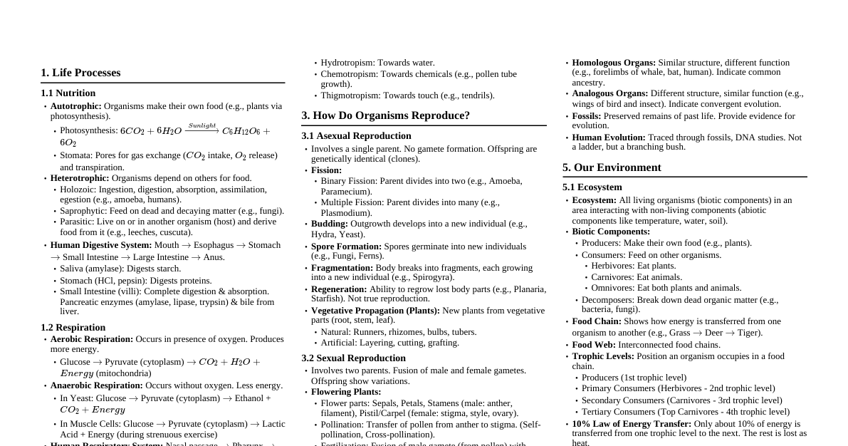
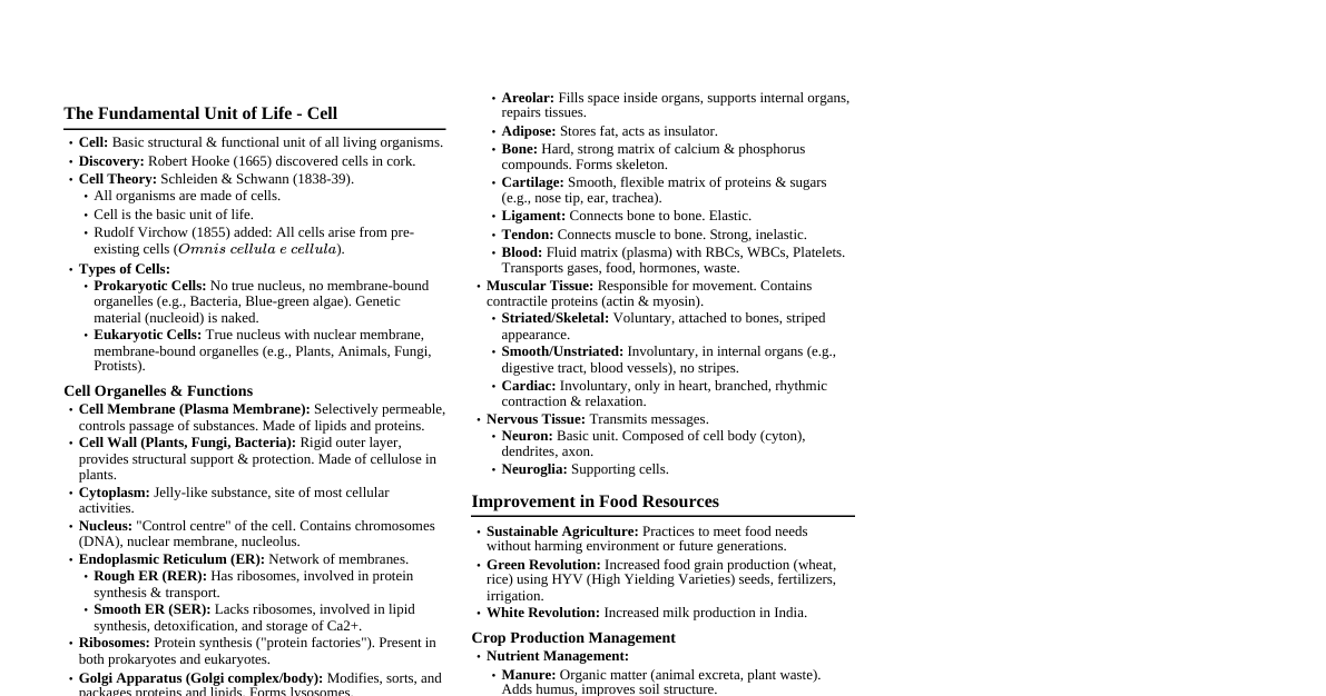
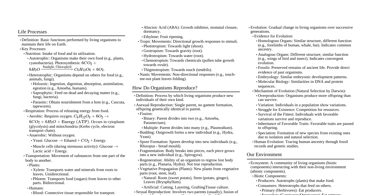
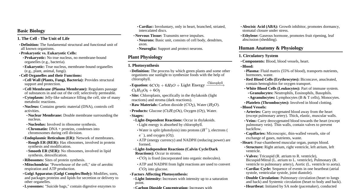
### Chapter 5: The Fundamental Unit of Life - The Cell (Page 1) #### 1. Introduction & Discovery - **Definition:** The fundamental structural and functional unit of all living organisms. All life functions occur within cells. - **Discovery Timeline:** - **Robert Hooke (1665):** First observed 'cells' (dead cork cells) and coined the term. - **Anton von Leeuwenhoek (1674):** First to observe living cells (bacteria, protozoa) in pond water. - **Robert Brown (1831):** Discovered the nucleus within the cell. - **Purkinje (1839):** Coined the term 'protoplasm' for the fluid substance of the cell. - **Schleiden (1838) & Schwann (1839):** Proposed the initial Cell Theory. - **Rudolf Virchow (1855):** Added the crucial concept *Omnis cellula e cellula* (All cells arise from pre-existing cells). #### 2. The Cell Theory 1. All living organisms are composed of cells and their products. 2. The cell is the basic structural and functional unit of life. 3. All cells arise from pre-existing cells through cell division. #### 3. Types of Cells: Prokaryotic vs. Eukaryotic Cells are broadly classified based on the presence or absence of a true nucleus and membrane-bound organelles. ```mermaid graph TD A[Cell Types] --> B{Prokaryotic Cells} A --> C{Eukaryotic Cells} B --> B1[No true nucleus (nucleoid region)] B --> B2[No membrane-bound organelles] B --> B3[Smaller (1-10 µm)] B --> B4[Circular DNA] B --> B5[Examples: Bacteria, Cyanobacteria] C --> C1[True nucleus with nuclear membrane] C --> C2[Membrane-bound organelles (ER, Golgi, Mitochondria, etc.)] C --> C3[Larger (5-100 µm)] C --> C4[Linear DNA] C --> C5[Examples: Plants, Animals, Fungi, Protists] ``` #### 4. Major Cell Components & Organelles ##### 4.1. Plasma Membrane (Cell Membrane) - **Description:** The outermost living boundary of the cell. It's a dynamic, flexible, and selectively permeable barrier. - **Structure:** Composed of a bi-lipid layer (phospholipids) with proteins embedded or attached (Fluid Mosaic Model). - **Function:** - Regulates the movement of substances into and out of the cell (selective permeability). - Maintains cell shape and integrity. - Receives external signals. - **Movement Across Membrane:** - **Diffusion:** Movement of substances from high to low concentration (e.g., gases). - **Osmosis:** Movement of water across a semi-permeable membrane from high to low water potential. - **Hypertonic solution:** Cell loses water, shrinks (plasmolysis in plants). - **Hypotonic solution:** Cell gains water, swells, may burst (animal cell) or become turgid (plant cell). - **Isotonic solution:** No net movement of water. - **Active Transport:** Movement of substances against their concentration gradient, requiring energy (ATP). ##### 4.2. Cell Wall (Present in Plant Cells, Fungi, Bacteria) - **Description:** A rigid, non-living outer layer found external to the plasma membrane. - **Structure:** Primarily composed of cellulose in plants, chitin in fungi, and peptidoglycan in bacteria. - **Function:** - Provides structural support and protection to the plant cell. - Prevents excessive water uptake (osmotic lysis) by forming a pressure against the swollen protoplast. - Maintains cell shape. - **Figure:** `(Imagine a diagram showing a thick outer cell wall surrounding the plasma membrane)` ##### 4.3. Cytoplasm - **Description:** The jelly-like, semi-fluid substance filling the space within the cell membrane, surrounding the nucleus and organelles. - **Components:** - **Cytosol:** The aqueous component of cytoplasm, where various metabolic reactions occur. - **Cell Organelles:** Membrane-bound structures with specific functions. - **Function:** Site of many cellular processes (e.g., glycolysis). ##### 4.4. Nucleus (The Control Center) - **Description:** The largest, most prominent organelle in eukaryotic cells, containing the cell's genetic material. - **Structure:** - **Nuclear Envelope:** A double membrane enclosing the nucleus, perforated by **nuclear pores** for transport. - **Nucleoplasm:** The fluid inside the nucleus. - **Chromatin:** A network of thread-like structures composed of DNA and associated proteins (histones). During cell division, chromatin condenses to form discrete **chromosomes**. - **Nucleolus:** A dense, spherical structure within the nucleus, responsible for ribosome synthesis. - **Function:** - Controls all cellular activities (growth, metabolism, protein synthesis). - Stores and protects the cell's genetic information (DNA). - Essential for cell reproduction and heredity. - **Figure:** `(Imagine a diagram of a nucleus with a double membrane, nuclear pores, chromatin, and nucleolus)` ##### 4.5. Endoplasmic Reticulum (ER) - **Description:** A vast network of membrane-bound tubules and flattened sacs (cisternae) extending from the nuclear envelope throughout the cytoplasm. - **Types & Function:** - **Rough ER (RER):** Studded with ribosomes. Primarily involved in the synthesis, folding, modification, and transport of proteins destined for secretion or insertion into membranes. - **Smooth ER (SER):** Lacks ribosomes. Involved in lipid synthesis (e.g., steroids), detoxification of drugs and poisons, and storage of calcium ions. - **Figure:** `(Imagine a diagram showing RER with ribosomes and SER as interconnected tubules)` ##### 4.6. Ribosomes - **Description:** Tiny, granular structures, either free in the cytoplasm or attached to the RER. - **Structure:** Composed of ribosomal RNA (rRNA) and proteins. Prokaryotic ribosomes are 70S, eukaryotic are 80S. - **Function:** Sites of protein synthesis (translating mRNA into protein sequences) – often called "protein factories." ##### 4.7. Golgi Apparatus (Golgi Complex/Body) - **Description:** A stack of flattened, membrane-bound sacs (cisternae) and associated vesicles. - **Function:** - Modifies, sorts, and packages proteins and lipids synthesized in the ER into vesicles for secretion or delivery to other organelles. - Involved in the formation of lysosomes and peroxisomes. - Plays a role in complex carbohydrate synthesis (e.g., cell wall components in plants). - **Figure:** `(Imagine a diagram showing stacks of flattened sacs with vesicles budding off)` ##### 4.8. Lysosomes (Waste Disposal System / Suicidal Bags) - **Description:** Small, spherical, membrane-bound sacs containing powerful hydrolytic (digestive) enzymes. - **Function:** - Breakdown of waste materials, cellular debris, worn-out organelles, and foreign invaders (e.g., bacteria). - Involved in programmed cell death (apoptosis), hence "suicidal bags." - **Figure:** `(Imagine a small, spherical sac with digestive enzymes inside)` ##### 4.9. Mitochondria (Powerhouse of the Cell) - **Description:** Rod-shaped or oval-shaped organelles responsible for cellular respiration. - **Structure:** - **Outer Membrane:** Smooth and permeable. - **Inner Membrane:** Highly folded into finger-like projections called **cristae**, increasing surface area. - **Matrix:** The fluid-filled space within the inner membrane, containing enzymes, ribosomes, and mitochondrial DNA. - **Function:** - Site of aerobic cellular respiration, where glucose is broken down to produce ATP (Adenosine Triphosphate) – the cell's energy currency. - **Figure:** `(Imagine an oval-shaped organelle with a smooth outer membrane and folded inner membrane (cristae)) ` ##### 4.10. Plastids (Present in Plant Cells & Algae) - **Description:** Large, double-membraned organelles found in plant cells. - **Types & Function:** - **Chloroplasts:** Green plastids containing chlorophyll. Site of photosynthesis (converts light energy into chemical energy). - **Structure:** Double membrane, stroma (fluid matrix), and stacks of thylakoids called grana. - **Chromoplasts:** Contain colored pigments (e.g., red in tomatoes, yellow in flower petals), responsible for non-green colors. - **Leucoplasts:** Colorless plastids primarily involved in storage (e.g., amyloplasts store starch, elaioplasts store oils, aleuroplasts store proteins). - **Figure:** `(Imagine a chloroplast with outer/inner membranes, stroma, and grana stacks)` ##### 4.11. Vacuoles (Storage Sacs) - **Description:** Membrane-bound sacs that vary greatly in size and function. - **Function:** - **Plants:** Typically a single, large central vacuole (up to 90% of cell volume). Stores water, nutrients, waste products, and maintains **turgor pressure** against the cell wall, providing support. - **Animals:** Small, temporary, or absent. May store water, food, or waste. - **Protists (e.g., Amoeba):** Food vacuoles for digestion, contractile vacuoles for osmoregulation. - **Figure:** `(Imagine a large central vacuole pushing the nucleus to the side in a plant cell, and small vesicles in an animal cell)` ##### 4.12. Centrosome (Present in Animal Cells & Lower Plants) - **Description:** An organelle located near the nucleus, containing two cylindrical structures called **centrioles**. - **Function:** Plays a crucial role in cell division by organizing the spindle fibers that separate chromosomes. #### 5. Plant Cell vs. Animal Cell ```mermaid graph TD A[Cell Types] --> B(Plant Cell) A --> C(Animal Cell) B --> B1[Cell Wall: Present] B --> B2[Chloroplasts: Present] B --> B3[Vacuole: Large, central, permanent] B --> B4[Centrosome: Absent (except lower plants)] B --> B5[Shape: Fixed, rectangular/square] B --> B6[Lysosomes: Rare or absent] C --> C1[Cell Wall: Absent] C --> C2[Chloroplasts: Absent] C --> C3[Vacuole: Small, temporary, or absent] C --> C4[Centrosome: Present] C --> C5[Shape: Irregular/round] C --> C6[Lysosomes: Present] ``` - **Figure: Typical Animal Cell** - **Figure: Typical Plant Cell** ### Chapter 6: Tissues (Page 2) #### 1. Introduction to Tissues - **Definition:** A group of cells that are similar in origin, structure, and function, specialized to perform a particular task. - **Importance:** - **Division of Labor:** Tissues allow for specialized functions, leading to increased efficiency in multicellular organisms. - **Higher Organization:** Enables the formation of organs and organ systems, crucial for complex life forms. - **Levels of Organization:** ```mermaid graph LR A[Cells] --> B[Tissues] B --> C[Organs] C --> D[Organ Systems] D --> E[Organism] ``` #### 2. Plant Tissues ##### 2.1. Meristematic Tissues (Growth Tissues) - **Characteristics:** Actively dividing cells, small, dense cytoplasm, prominent nuclei, thin cell walls, little or no vacuoles. - **Function:** Responsible for growth in plants, producing new cells. - **Types & Location:** 1. **Apical Meristem:** - **Location:** Found at the growing tips of roots and stems. - **Function:** Increases the length of the plant (primary growth). - **Example:** Growth of root tip and shoot tip. 2. **Lateral Meristem (Cambium):** - **Location:** Found along the sides of stems and roots, responsible for increasing girth. - **Function:** Increases the width or thickness of the plant (secondary growth). - **Example:** Vascular cambium in dicot stems. 3. **Intercalary Meristem:** - **Location:** Found at the base of leaves or internodes (e.g., grasses). - **Function:** Increases the length of organs, allows for regrowth of parts removed by grazing. - **Example:** Growth of grass blades after mowing. - **Figure:** `(Imagine a diagram of a plant showing apical meristem at shoot/root tips, and lateral meristem in stem circumference)` ##### 2.2. Permanent Tissues (Specialized Tissues) - **Characteristics:** Cells that have lost the ability to divide, have taken a permanent shape, size, and function through differentiation. - **Origin:** Formed from meristematic tissues. ###### 2.2.1. Simple Permanent Tissues (Composed of one type of cell) 1. **Parenchyma:** - **Structure:** Thin-walled, living cells, often loosely packed with large intercellular spaces, typically isodiametric (spherical/oval), large central vacuole. - **Function:** - **Storage:** Stores food (starch, oils, proteins) and water. - **Photosynthesis:** If it contains chloroplasts (chlorenchyma, e.g., in leaves). - **Buoyancy:** Aerenchyma (parenchyma with large air cavities) provides buoyancy to aquatic plants. - **Turgidity:** Provides structural support when turgid. - **Location:** Cortex of roots, pith of stems, pulp of fruits, mesophyll of leaves. - **Figure:** `(Diagram: Loosely packed, roundish cells with thin walls)` 2. **Collenchyma:** - **Structure:** Living cells, elongated, irregularly thickened at the corners (due to deposition of pectin and cellulose), very little intercellular space. - **Function:** Provides mechanical support and flexibility to young stems and petioles, allowing bending without breaking. - **Location:** Below the epidermis of dicot stems, petiole of leaves. - **Figure:** `(Diagram: Cells with noticeably thickened corners)` 3. **Sclerenchyma:** - **Structure:** Dead cells, long and narrow (fibers) or short and irregular (sclereids), with thick and lignified (woody) cell walls, no internal protoplasm. - **Function:** Provides strength, rigidity, and hardness to plant parts, protecting them from mechanical stress. - **Types:** - **Fibers:** Long, pointed cells (e.g., in jute, hemp, coconut husk). - **Sclereids (Stone cells):** Short, spherical, or oval cells (e.g., hard covering of nuts, pulp of guava/pear). - **Location:** Around vascular bundles, veins of leaves, hard coverings of seeds and nuts. - **Figure:** `(Diagram: Thick-walled, tightly packed cells, some elongated, others irregular)` ###### 2.2.2. Complex Permanent Tissues (Composed of more than one type of cell, working together) 1. **Xylem (Water-conducting tissue):** - **Definition:** The primary water-conducting tissue in plants. - **Components:** - **Tracheids:** Elongated, tapering dead cells with pits. - **Vessels:** Tube-like structures formed by rows of dead cells placed end-to-end, forming a continuous pipe. - **Xylem Parenchyma:** Living cells, stores food. - **Xylem Fibers:** Dead, sclerenchymatous cells providing mechanical support. - **Function:** Transports water and dissolved minerals from roots to all parts of the plant. Also provides mechanical strength. - **Direction:** Unidirectional (upwards). - **Figure:** `(Diagram: Cross-section showing large vessel elements and smaller tracheids)` 2. **Phloem (Food-conducting tissue):** - **Definition:** The primary food-conducting tissue in plants. - **Components:** - **Sieve Tubes:** Living, elongated cells with perforated end walls (sieve plates) for transport. - **Companion Cells:** Living cells, associated with sieve tubes, help in their metabolic activities. - **Phloem Parenchyma:** Living cells, stores food. - **Phloem Fibers:** Sclerenchymatous cells (dead), provide support. - **Function:** Transports prepared food (sugars, mainly sucrose) from leaves (site of photosynthesis) to other parts of the plant where it is needed for growth or storage. - **Direction:** Bidirectional. - **Figure:** `(Diagram: Cross-section showing sieve tubes with sieve plates and adjacent companion cells)` #### 3. Animal Tissues ##### 3.1. Epithelial Tissue (Covering & Lining Tissue) - **Characteristics:** Cells are tightly packed, forming continuous sheets with very little intercellular matrix. Cells rest on a non-cellular basement membrane. - **Function:** Protection, secretion, absorption, excretion, filtration. - **Types & Location:** 1. **Squamous Epithelium:** - **Structure:** Extremely thin, flat, and irregular cells, forming a delicate lining. - **Function:** Forms a permeable surface for diffusion and filtration. - **Location:** Lining of blood vessels (endothelium), lung alveoli, oesophagus, mouth. - **Figure:** `(Diagram: A single layer of flat, scale-like cells, like tiles on a floor)` 2. **Cuboidal Epithelium:** - **Structure:** Cube-shaped cells, often with central spherical nuclei. - **Function:** Secretion and absorption; provides mechanical support. - **Location:** Lining of kidney tubules, ducts of salivary glands, thyroid follicles. - **Figure:** `(Diagram: A single layer of cube-shaped cells)` 3. **Columnar Epithelium:** - **Structure:** Tall, pillar-like cells, with elongated nuclei usually near the base. - **Function:** Secretion (e.g., mucus by goblet cells) and absorption. - **Location:** Lining of stomach, intestine, gall bladder. - **Figure:** `(Diagram: A single layer of tall, rectangular cells)` 4. **Ciliated Columnar Epithelium:** - **Structure:** Columnar cells with hair-like projections called **cilia** on their free surface. - **Function:** Cilia beat in a coordinated manner to move substances (e.g., mucus, ova) in a specific direction. - **Location:** Lining of respiratory tract (trachea, bronchi), Fallopian tubes. - **Figure:** `(Diagram: Columnar cells with cilia on their apical surface)` 5. **Stratified Squamous Epithelium:** - **Structure:** Multiple layers of squamous cells. The outermost layers are flattened, while deeper layers are cuboidal. - **Function:** Provides protection against wear and tear, abrasion, and drying. - **Location:** Skin (keratinized), lining of oesophagus, oral cavity, vagina. - **Figure:** `(Diagram: Multiple layers of cells, with flat cells at the top and cuboidal cells at the bottom)` 6. **Glandular Epithelium:** Modified epithelial cells that specialize in secreting substances (e.g., sweat glands, endocrine glands). ##### 3.2. Connective Tissue (Binding & Supporting Tissue) - **Characteristics:** Cells are loosely packed and embedded in a large amount of intercellular matrix (which can be jelly-like, fluid, dense, or rigid). The matrix is secreted by the cells themselves. - **Function:** Connects various organs, provides support, protection, insulation, and transport. - **Types & Location:** 1. **Areolar Connective Tissue:** - **Structure:** Loose and irregular, contains various cells (fibroblasts, macrophages, mast cells) and fibers (collagen, elastin) in a jelly-like matrix. - **Function:** Fills spaces inside organs, supports internal organs, helps in tissue repair, attaches skin to underlying muscles. - **Location:** Found between skin and muscles, around blood vessels and nerves, in bone marrow. - **Figure:** `(Diagram: A loose network of fibers and scattered cells)` 2. **Adipose Tissue:** - **Structure:** Composed of specialized cells called adipocytes, which are filled with large fat globules, pushing the nucleus to the periphery. - **Function:** Stores fat (energy reserve), acts as an insulator against cold, cushions organs (e.g., kidneys, eyeballs). - **Location:** Below the skin, between internal organs, around heart. - **Figure:** `(Diagram: Large, vacuolated cells, with nucleus pushed to the side)` 3. **Bone:** - **Structure:** Hard, rigid, and porous matrix composed of calcium and phosphorus compounds (mineral salts) and collagen fibers. Bone cells (osteocytes) are present in small cavities called lacunae. - **Function:** Forms the skeletal framework, provides structural support, protects vital organs, anchors muscles, stores minerals. - **Location:** The entire skeleton. - **Figure:** `(Diagram: Concentric rings (lamellae) of matrix around a central Haversian canal, with osteocytes in lacunae)` 4. **Cartilage:** - **Structure:** Firm, flexible, and elastic matrix (composed of proteins and sugars). Cartilage cells (chondrocytes) are enclosed in small cavities within the matrix. - **Function:** Provides flexibility and support, cushions bones at joints, absorbs shock. - **Location:** Tip of nose, outer ear, trachea, larynx, ends of long bones, intervertebral discs. - **Figure:** `(Diagram: Chondrocytes in lacunae within a smooth, elastic matrix)` 5. **Tendons:** - **Structure:** Dense regular connective tissue, made of white fibrous collagen fibers arranged in parallel bundles. Possesses great strength but limited flexibility. - **Function:** Connects muscles to bones. - **Figure:** `(Diagram: Tightly packed, parallel collagen fibers)` 6. **Ligaments:** - **Structure:** Dense regular connective tissue, very elastic, contains more yellow elastic fibers than tendons. Strong but flexible. - **Function:** Connects bone to bone, holding joints together. - **Figure:** `(Diagram: Less regularly arranged elastic fibers compared to tendons)` 7. **Blood (Fluid Connective Tissue):** - **Structure:** Fluid matrix called **plasma** (water, proteins, salts, hormones, waste products) in which various blood cells are suspended. - **Components:** - **Red Blood Cells (RBCs / Erythrocytes):** Biconcave disc-shaped, anucleated (in mammals), contain hemoglobin for oxygen transport. - **White Blood Cells (WBCs / Leukocytes):** Colorless, nucleated, involved in immune response (fight infection). - **Platelets (Thrombocytes):** Cell fragments, help in blood clotting. - **Function:** Transports gases (O2, CO2), digested food, hormones, and waste products throughout the body. Regulates body temperature. - **Figure:** `(Diagram: Different blood cells (RBC, WBC, Platelets) floating in plasma)` ##### 3.3. Muscular Tissue (Movement Tissue) - **Characteristics:** Composed of elongated cells called muscle fibers, which contain contractile proteins (actin and myosin) that allow for movement. - **Function:** Responsible for all types of movement in the body. - **Types & Control:** 1. **Striated (Skeletal/Voluntary) Muscle:** - **Control:** Voluntary (under conscious control). - **Structure:** Long, cylindrical, unbranched fibers, multinucleated, show distinct light and dark bands (striations). - **Function:** Movement of bones and body parts. - **Location:** Attached to bones (e.g., biceps, triceps, muscles of limbs). - **Figure:** `(Diagram: Long, unbranched, striped fibers with multiple nuclei)` 2. **Smooth (Unstriated/Involuntary) Muscle:** - **Control:** Involuntary (not under conscious control). - **Structure:** Spindle-shaped (tapering at ends), uninucleated, no striations. - **Function:** Controls involuntary movements (e.g., movement of food in the alimentary canal, contraction of blood vessels, constriction of pupils). - **Location:** Walls of internal organs like stomach, intestine, blood vessels, iris of the eye, uterus, urinary bladder. - **Figure:** `(Diagram: Spindle-shaped cells without stripes, single nucleus)` 3. **Cardiac Muscle:** - **Control:** Involuntary. - **Structure:** Cylindrical, branched fibers, uninucleated, show faint striations, interconnected by specialized junctions called **intercalated discs**. - **Function:** Rhythmic contraction and relaxation of the heart throughout life, pumping blood. - **Location:** Wall of the heart only. - **Figure:** `(Diagram: Branched, faintly striped cells with intercalated discs and single nucleus)` ##### 3.4. Nervous Tissue (Communication Tissue) - **Characteristics:** Highly specialized for transmitting electrical signals (nerve impulses) very rapidly from one part of the body to another. - **Components:** 1. **Neurons (Nerve Cells):** - **Structure:** - **Cell Body (Soma/Cyton):** Contains the nucleus and most of the cytoplasm. - **Dendrites:** Short, branched extensions that receive nerve impulses from other neurons. - **Axon:** A single, long extension that transmits nerve impulses away from the cell body to other neurons, muscles, or glands. May be covered by a myelin sheath. - **Function:** Transmit electrical signals (nerve impulses) throughout the body. - **Figure:** `(Diagram: Classic neuron structure with dendrites, cell body, and a long axon)` 2. **Neuroglia (Glial Cells):** - **Function:** Support, protect, and nourish neurons; do not transmit nerve impulses themselves. - **Location:** Brain, spinal cord, and nerves throughout the body. #### 4. Flowchart: Animal Tissue Classification ```mermaid graph TD A[Animal Tissues] --> B{Epithelial Tissue} A --> C{Connective Tissue} A --> D{Muscular Tissue} A --> E{Nervous Tissue} B --> B1[Squamous: flat, diffusion (lungs)] B --> B2[Cuboidal: cube, secretion/absorption (kidney tubules)] B --> B3[Columnar: tall, absorption/secretion (intestine)] B --> B4[Ciliated Columnar: cilia, movement (respiratory tract)] B --> B5[Stratified Squamous: layers, protection (skin)] C --> C1[Areolar: loose, fills space (under skin)] C --> C2[Adipose: fat storage, insulation (below skin)] C --> C3[Bone: rigid, support (skeleton)] C --> C4[Cartilage: flexible, cushioning (nose, ear)] C --> C5[Tendons: muscle to bone, strong] C --> C6[Ligaments: bone to bone, elastic] C --> C7[Blood: fluid, transport (whole body)] D --> D1[Striated (Skeletal): voluntary, attached to bone] D --> D2[Smooth (Involuntary): involuntary, internal organs] D --> D3[Cardiac: involuntary, heart only] E --> E1[Neurons: transmit impulses] E --> E2[Neuroglia: support neurons] ```