CBSE Class 12 Biology Practicals
Cheatsheet Content
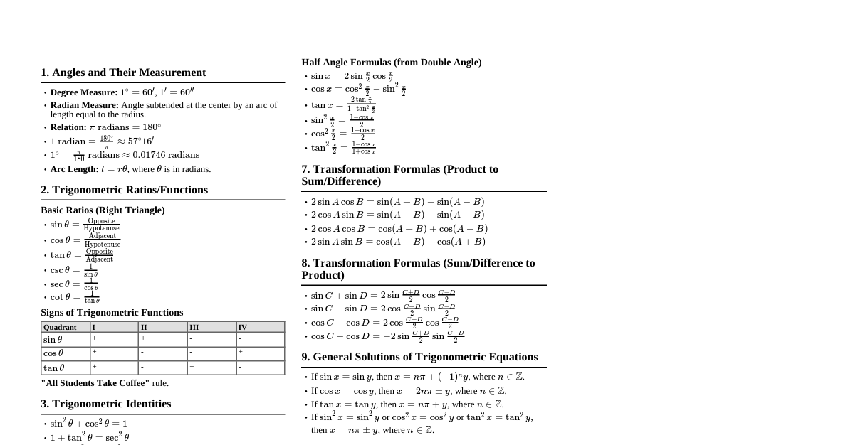
1. Study of Plant Population Density by Quadrat Method Aim: To estimate plant population density using the quadrat method. Principle: Density is the number of individuals of a species per unit area. Quadrat is a sampling unit. Materials: Quadrat (e.g., $1m \times 1m$ square), measuring tape, string, pegs, notebook, pen. Procedure: Select a study site. Place quadrats randomly or systematically within the site. Count the number of individuals of each plant species within each quadrat. Record observations in a table. Calculations: Density (D) = $\frac{\text{Total number of individuals of a species in all quadrats}}{\text{Total number of quadrats studied} \times \text{Area of one quadrat}}$ Population Density = $\frac{\text{Total number of individuals of a species}}{\text{Total sampling area}}$ Precautions: Random placement of quadrats, accurate counting, proper identification of species. 2. Study of Plant Population Frequency by Quadrat Method Aim: To estimate plant population frequency using the quadrat method. Principle: Frequency is the percentage of quadrats in which a given species occurs. It indicates distribution pattern. Materials: Same as for population density. Procedure: Lay quadrats as described for density. Note the presence or absence of each species in each quadrat. Record observations. Calculations: Frequency (F) % = $\frac{\text{Number of quadrats in which the species occurred}}{\text{Total number of quadrats studied}} \times 100$ Interpretation: High frequency indicates wide distribution. 3. Study of Water Holding Capacity of Soils Aim: To compare the water holding capacity of different soil samples (e.g., sandy, loamy, clayey). Principle: Water holding capacity is the amount of water retained by a soil sample against gravity. Materials: Funnels, filter paper, measuring cylinders, different soil samples, distilled water, weighing balance. Procedure: Place filter paper in funnels. Add known weight (e.g., 50g) of dry soil to each funnel. Place funnels over measuring cylinders. Pour water slowly until it starts dripping. Allow to drain completely. Measure the volume of water collected in the cylinder. Weigh the wet soil sample. Calculations: Volume of water absorbed by soil = Volume of water added - Volume of water drained. Water holding capacity (%) = $\frac{\text{Weight of water absorbed}}{\text{Weight of dry soil}} \times 100$ Weight of water absorbed = Volume of water absorbed (since density of water is $1 g/mL$). Observation: Clayey soil has highest, sandy soil has lowest water holding capacity. 4. Study of Presence of Suspended Particulate Matter in Air Aim: To study the presence of suspended particulate matter (SPM) in air at different sites. Principle: Particulate matter gets trapped on filter paper when air is drawn through it. Materials: Suction pump/vacuum cleaner, filter paper (pre-weighed), petri dishes, weighing balance, stopwatch. Procedure: Weigh a clean, dry filter paper ($W_1$). Attach it to the suction pump outlet. Operate the pump for a fixed duration (e.g., 30 mins) at a specific site. Remove and dry the filter paper. Weigh the filter paper again ($W_2$). Repeat for different sites (e.g., roadside, residential area). Calculations: Weight of SPM = $W_2 - W_1$ Compare SPM levels across different sites. Precautions: Ensure airtight connections, use calibrated balance, control for environmental factors. 5. Study of Presence of Microbes in Water Samples Aim: To study the presence of microbes in different water samples (pond water, tap water, sewage water). Principle: Microbes grow and form colonies on suitable nutrient media. Materials: Sterilized petri dishes, nutrient agar medium, water samples, autoclave, incubator, inoculating loop, spirit lamp. Procedure: Prepare and sterilize nutrient agar medium. Pour agar into sterile petri dishes and allow to solidify (control plate). Take different water samples. Using a sterile inoculating loop, streak a loopful of each water sample onto separate agar plates. Incubate plates (e.g., at $37^\circ C$) for 24-48 hours. Observe and count the number of microbial colonies. Observation: Sewage water will show the highest microbial growth, tap water the least. Precautions: Maintain aseptic conditions, proper sterilization, careful handling of cultures. 6. Preparation of Temporary Mount of Onion Root Tip for Mitosis Aim: To study different stages of mitosis in onion root tip cells. Principle: Meristematic cells at the root tip undergo continuous cell division (mitosis). Materials: Onion root tips (pre-treated with fixative like Carnoy's fluid and hydrolyzed in HCl), acetocarmine stain, slide, coverslip, blotting paper, microscope. Procedure: Take a hydrolyzed root tip (softened). Place it on a clean slide. Add a drop of acetocarmine stain. Macerate the tip gently with a needle. Place a coverslip and tap gently to spread cells (squash preparation). Observe under the microscope. Observation: Identify interphase, prophase, metaphase, anaphase, telophase. Key features: Interphase: Nucleus visible, no distinct chromosomes. Prophase: Chromosomes condense, nuclear envelope disappears. Metaphase: Chromosomes align at equatorial plate. Anaphase: Sister chromatids separate and move to opposite poles. Telophase: Chromosomes decondense, nuclear envelope reforms, cytokinesis begins. 7. Study of Pollen Germination on a Slide Aim: To study pollen germination on a slide. Principle: Pollen grains absorb moisture and nutrients from a suitable medium, leading to the formation of a pollen tube. Materials: Fresh pollen grains (e.g., Balsam, Petunia, Lily), cavity slide, coverslip, sucrose solution (10%), boric acid, distilled water, microscope. Procedure: Prepare a germination medium (e.g., 10% sucrose solution with a trace of boric acid). Place a drop of medium on a cavity slide. Dust fresh pollen grains onto the drop. Place a coverslip. Incubate at room temperature for 15-30 minutes. Observe under the microscope. Observation: Observe swollen pollen grains and the emergence and growth of pollen tubes. Significance: Essential for fertilization in plants. 8. Study of Meiosis in Onion Bud Cells or Grasshopper Testis Aim: To study different stages of meiosis. Principle: Meiosis is reductional division occurring in germ cells, leading to haploid gametes. Materials: Onion flower buds (pre-treated), acetocarmine stain, slide, coverslip, microscope. Procedure: Take an anther from a flower bud. Place it on a slide, add a drop of stain. Macerate and squash gently. Observe under the microscope. Observation: Identify Meiosis I (Prophase I, Metaphase I, Anaphase I, Telophase I) and Meiosis II (Prophase II, Metaphase II, Anaphase II, Telophase II). Key features (Meiosis I): Prophase I: Longest, includes leptotene, zygotene (pairing/synapsis), pachytene (crossing over), diplotene, diakinesis. Metaphase I: Homologous pairs align at the metaphase plate. Anaphase I: Homologous chromosomes separate. Telophase I: Two haploid cells form. Key features (Meiosis II): Similar to mitosis, but starts with haploid cells. Sister chromatids separate. 9. Study of DNA Isolation from Plant Material (e.g., Spinach, Papaya) Aim: To isolate DNA from plant material. Principle: DNA can be extracted by breaking cell membranes, denaturing proteins, and precipitating DNA. Materials: Fresh plant material (e.g., spinach leaves), mortar and pestle, beaker, test tubes, distilled water, common salt (NaCl), liquid detergent, chilled ethanol/isopropanol. Procedure: Grind plant material with a pinch of salt and a small amount of distilled water to break cell walls. Add liquid detergent (e.g., dish soap) to lyse cell membranes and nuclear envelope. Incubate mixture at $60^\circ C$ for 10-15 min (helps denature enzymes). Filter the mixture through a muslin cloth into a clean test tube. Carefully pour chilled ethanol/isopropanol down the side of the test tube. DNA precipitates as white, fibrous strands at the interface. Spool the DNA using a glass rod. Role of chemicals: Salt: Neutralizes charge on DNA, aids precipitation. Detergent: Emulsifies lipids in cell membranes, releases DNA. Ethanol: DNA is insoluble in chilled ethanol, causing it to precipitate. 10. Study of Mendelian Inheritance using Seeds of Different Colours/Textures Aim: To demonstrate Mendelian inheritance (Monohybrid/Dihybrid Cross) using seeds. Principle: Demonstrates dominance, segregation, and independent assortment. Materials: Seeds of different colours/textures (e.g., yellow/green peas, round/wrinkled peas), trays, counter. Procedure (Monohybrid Cross): Take $F_1$ generation (all dominant phenotype, e.g., all yellow). Assume $F_1$ self-pollinates to produce $F_2$. Randomly mix $F_1$ seeds representing heterozygotes (e.g., $Yy$). Randomly pick a large number of 'gametes' ($Y$ or $y$) and combine them to form $F_2$ genotypes ($YY, Yy, yy$). Count the number of seeds corresponding to dominant and recessive phenotypes. Expected Ratios: Monohybrid (phenotypic): 3 (Dominant) : 1 (Recessive) Monohybrid (genotypic): 1 ($AA$) : 2 ($Aa$) : 1 ($aa$) Dihybrid (phenotypic): 9 ($A\_B\_$) : 3 ($A\_bb$) : 3 ($aaB\_$) : 1 ($aabb$) Conclusion: Observed ratios should approximate theoretical Mendelian ratios. 11. Study of Different Types of Pollination in Flowers Aim: To observe and identify different types of pollination in flowers. Principle: Pollination is the transfer of pollen grains from anther to stigma. Can be self or cross-pollination. Materials: Various flowers (e.g., Hibiscus, Salvia, Maize, Papaya), hand lens, dissecting needle. Procedure: Observe floral parts (anthers, stigma, petals, nectaries). Note arrangement of sexual organs. Identify adaptations for different pollinators (e.g., large petals for insects, feathery stigma for wind). Types of Pollination: Anemophily (Wind): Small, inconspicuous flowers, no scent/nectar, large feathery stigma, dusty pollen (e.g., Maize, Grasses). Entomophily (Insect): Large, brightly coloured/scented flowers, nectar guides, sticky pollen, prominent stigma (e.g., Hibiscus, Salvia). Hydrophily (Water): Rare, small flowers, unwettable pollen (e.g., Vallisneria). Ornithophily (Bird): Brightly coloured, tubular flowers, abundant nectar (e.g., Butea). Self-pollination: Transfer of pollen within the same flower or between flowers on the same plant. Cross-pollination: Transfer of pollen between flowers of different plants of the same species. 12. Study of Dissection of Flowers (e.g., Hibiscus) Aim: To study the morphology of a flower by dissection. Principle: Understanding floral parts and their arrangement. Materials: Fresh flower (e.g., Hibiscus), dissecting tray, forceps, dissecting needles, scalpel, hand lens, drawing sheet. Procedure: Observe external features: pedicel, bracts, calyx, corolla. Carefully remove sepals and petals. Observe androecium (stamens: anther, filament). Observe gynoecium (pistil: stigma, style, ovary). Cut a transverse and longitudinal section of the ovary to observe ovules. Draw labeled diagrams of all parts. Key Parts of Hibiscus: Calyx: 5 fused sepals, epicalyx present. Corolla: 5 free petals. Androecium: Numerous stamens, monadelphous (fused filaments forming a staminal tube), anthers extrorse. Gynoecium: Pentacarpellary, syncarpous, superior ovary, many ovules in each locule, 5-lobed stigma. 13. Study of Homology and Analogy from Specimens/Charts Aim: To distinguish between homologous and analogous organs. Principle: Homology indicates common ancestry; analogy indicates convergent evolution. Materials: Charts/specimens of forelimbs of vertebrates (homologous) and wings of insects/birds (analogous). Homologous Organs: Definition: Organs with similar basic structure and embryonic origin but perform different functions. Example: Forelimbs of whale, bat, cheetah, human (all have humerus, radius, ulna, carpals, metacarpals, phalanges). Indicates: Divergent evolution from a common ancestor. Analogous Organs: Definition: Organs with different basic structure and embryonic origin but perform similar functions. Example: Wings of insect and wings of bird (insect wing is chitinous membrane, bird wing is bony skeleton covered with feathers). Indicates: Convergent evolution due to similar environmental pressures. 14. Study of Adaptations of Plants and Animals Aim: To identify morphological adaptations in organisms. Principle: Organisms develop specific features to survive in their habitats. Materials: Specimens/charts of xerophytes (e.g., Opuntia, Acacia), hydrophytes (e.g., Hydrilla, Eichhornia), animals (e.g., fish, frog, camel, bird). Xerophytes (Dry Habitats): Leaves: Reduced, spines (Opuntia), leathery, sunken stomata (Nerium), thick cuticle. Stem: Fleshy, flattened (phylloclade in Opuntia), green (photosynthetic). Roots: Extensive. Example: Opuntia, Acacia, Nerium. Hydrophytes (Aquatic Habitats): Submerged: Finely dissected leaves, no stomata, poor vascular tissue (Hydrilla). Floating: Large, broad leaves (water lily), air-filled petioles (Eichhornia), stomata on upper surface. Roots: Poorly developed or absent. Example: Hydrilla, Eichhornia, Lotus. Animal Adaptations: Fish (Aquatic): Streamlined body, gills for respiration, fins for movement, scales. Frog (Amphibian): Webbed feet, moist skin, camouflage, hibernation/aestivation. Camel (Desert): Hump for fat storage, thick fur, broad padded feet, ability to conserve water. Bird (Aerial): Streamlined body, hollow bones, wings, feathers. 15. Study of Permanent Slides of T.S. of Blastula, Gastrula Aim: To identify developmental stages from permanent slides. Principle: Early embryonic development involves cleavage, blastulation, and gastrulation. Materials: Permanent slides of blastula and gastrula (e.g., frog, chick). Observation: Blastula: Hollow, spherical structure. Outer layer of cells (blastomeres) enclosing a fluid-filled cavity (blastocoel). No increase in overall size from zygote. Gastrula: Formation of primary germ layers (ectoderm, mesoderm, endoderm). Blastocoel reduces or disappears. Formation of archenteron (primitive gut) and blastopore. Cells rearrange through morphogenetic movements. 16. Study of Permanent Slides of L.S. of Ovary, Testis Aim: To identify different stages of gamete formation. Principle: Oogenesis occurs in ovary, spermatogenesis in testis. Materials: Permanent slides of L.S. mammalian ovary and testis. Ovary (L.S.): Outer cortex: Contains ovarian follicles at various stages of development. Primordial follicles: Primary oocyte surrounded by a single layer of granulosa cells. Primary follicles: Primary oocyte surrounded by multiple layers of granulosa cells. Secondary follicles: Antrum (fluid-filled cavity) appears. Graafian follicle: Mature follicle, large antrum, eccentric oocyte surrounded by zona pellucida and corona radiata. Corpus luteum: Formed from ruptured Graafian follicle after ovulation. Medulla: Inner region, contains connective tissue, blood vessels, nerves. Testis (L.S.): Seminiferous tubules: Site of spermatogenesis. Germinal epithelium: Lines tubules, contains spermatogonia, primary spermatocytes, secondary spermatocytes, spermatids, spermatozoa. Sertoli cells: Nurse cells, provide nourishment to developing sperm. Interstitial (Leydig) cells: Located between tubules, secrete androgens.