Maxillary 1st Molar Cheatsheet
Cheatsheet Content
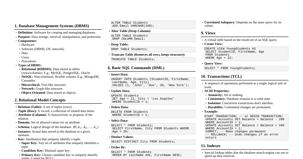
Maxillary First Molar (Tooth #3, #14) 1. Chronology Calcification begins: Birth Crown completed: 2.5 - 3 years Eruption: 6 - 7 years Root completed: 9 - 10 years 2. Dimensions (Average) Crown Length (cervical to cusp tip): 7.5 mm Root Length (buccal): 12.5 mm Mesiodistal Crown Diameter: 10 mm Faciolingual Crown Diameter: 11 mm Mesiodistal Diameter at Cervix: 8 mm Faciolingual Diameter at Cervix: 10 mm Curvature of Cervical Line (Mesial): 1 mm Curvature of Cervical Line (Distal): 0 mm 3. Morphology - General Characteristics Largest tooth in the maxillary arch. Often considered the "keystone" of the dental arch. Typical 4 cusps (MB, DB, ML, DL) and often a 5th cusp (Cusp of Carabelli). Three roots: Mesiobuccal, Distobuccal, Lingual (largest and longest). Rhomboidal or parallelogram occlusal outline. Morphology - Specific Aspects 1. Buccal Aspect Crown outline: Trapezoidal. Cusps: Mesiobuccal (MB) and Distobuccal (DB). MB cusp is broader and longer. Buccal groove: Divides the MB and DB cusps, often ends in a buccal pit. Cervical line: Slight curvature towards the root. Roots: Two buccal roots (MB and DB). MB root is broader, DB root is more pointed. Usually diverge, then converge. Space for Diagram: Buccal View 2. Lingual Aspect Crown outline: Trapezoidal. Cusps: Mesiolingual (ML) and Distolingual (DL). ML cusp is the largest and longest. DL cusp is the smallest. Cusp of Carabelli: Present on the mesiolingual surface of ML cusp in about 60% of cases. Lingual groove: Separates ML and DL cusps. Roots: Lingual root is the longest, largest, and most divergent, extending beyond the buccal roots. Space for Diagram: Lingual View 3. Mesial Aspect Crown outline: Trapezoidal. Cusps: ML cusp is the longest, followed by MB. Cervical line: Curves occlusally (1 mm). Contact area: Broad, located at the junction of the occlusal and middle thirds. Roots: MB and Lingual roots are visible. Lingual root is much wider and longer. Space for Diagram: Mesial View 4. Distal Aspect Crown outline: Trapezoidal. Cusps: DB cusp is visible, often shorter than MB. DL cusp is also visible. Cervical line: Straight or only slightly curved. Contact area: Smaller than mesial, located more cervically. Roots: All three roots are often visible (DB, MB, Lingual). Space for Diagram: Distal View 5. Occlusal Aspect Outline: Rhomboidal or parallelogram shape. Major Cusps: Mesiolingual (ML): Largest, longest, sharpest. Mesiobuccal (MB): Second largest. Distobuccal (DB): Third largest. Distolingual (DL): Smallest. Cusp of Carabelli: On ML cusp, if present. Ridges: Cusp Ridges: Mesial and distal ridges extending from each cusp tip. Triangular Ridges: Extend from cusp tips towards the central groove (e.g., MB triangular ridge, ML triangular ridge). Transverse Ridge: Formed by the union of the MB triangular ridge and the DL triangular ridge. Oblique Ridge: Unique to maxillary molars. Formed by the union of the ML triangular ridge and the DB triangular ridge. Runs from the ML cusp to the DB cusp. Marginal Ridges: Mesial marginal ridge (broader) and Distal marginal ridge. Grooves: Central Groove: Runs mesiodistally, connecting central pit to marginal ridges. Buccal Groove: Extends buccally from central groove, separating MB and DB cusps. Lingual Groove: Extends lingually from central groove, separating ML and DL cusps. Distal Oblique Groove: Often present, crosses the oblique ridge distally. Transverse Groove of Oblique Ridge: Sometimes crosses the oblique ridge. Supplemental Grooves: Numerous, irregular grooves on the occlusal surface. Pits: Central Pit: In the center of the occlusal surface, where central, buccal, and lingual grooves often meet. Mesial Triangular Fossa Pit: In the mesial triangular fossa. Distal Triangular Fossa Pit: In the distal triangular fossa. Buccal Pit: At the end of the buccal groove. Lingual Pit: At the end of the lingual groove (less common than buccal pit). Space for Diagram: Occlusal View 6. Contact Areas Mesial Contact: With maxillary 2nd premolar, occlusal/middle third junction, slightly buccal. Distal Contact: With maxillary 2nd molar, middle third, slightly buccal, more cervical. 7. Pulp Morphology Pulp Chamber: Corresponds to crown, 4 pulp horns (ML largest). Root Canals: 3 main canals. Lingual: Largest, round/oval. Distobuccal: Smallest, round/oval. Mesiobuccal: Often 2 canals (MB1, MB2), MB2 is common for missed canals. Accessory canals common (apical/furcation). Space for Diagram: Pulp Chamber and Root Canals Clinical Significance & Correlation 1. Occlusion & Function Keystone: Maintains arch integrity; loss impacts occlusion. Oblique Ridge: Key for crown strength and mastication. 2. Caries & Restorations Deep Pits/Fissures: High caries risk (central, buccal pits, grooves); need sealants. Cusp of Carabelli: Can have deep groove, prone to caries. Restorations: Complex anatomy requires precise carving. 3. Periodontal Health Divergent Roots: Challenging for scaling/root planing. Trifurcation: Common site for periodontal disease. 4. Endodontic Treatment MB2 Canal: Frequently present, critical to locate and treat to prevent failure. Pulp Chamber: Variable depth, careful access needed. Root Curvatures: Can complicate instrumentation (ledging/perforation risk). 5. Oral Surgery & Extraction Divergent Roots: Increases risk of root/tuberosity fracture during extraction. Maxillary Sinus Proximity: Risk of oro-antral communication or sinus involvement with periapical pathology. 6. Prosthodontics Crown Prep: Requires significant reduction due to cuspal/ridge anatomy. Abutment: Excellent for bridges/RPDs, but root divergence affects path of insertion.