Breathing & Gas Exchange (Class 12)
Cheatsheet Content
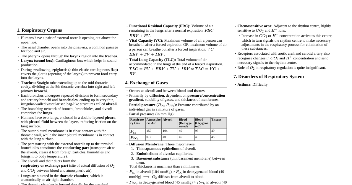
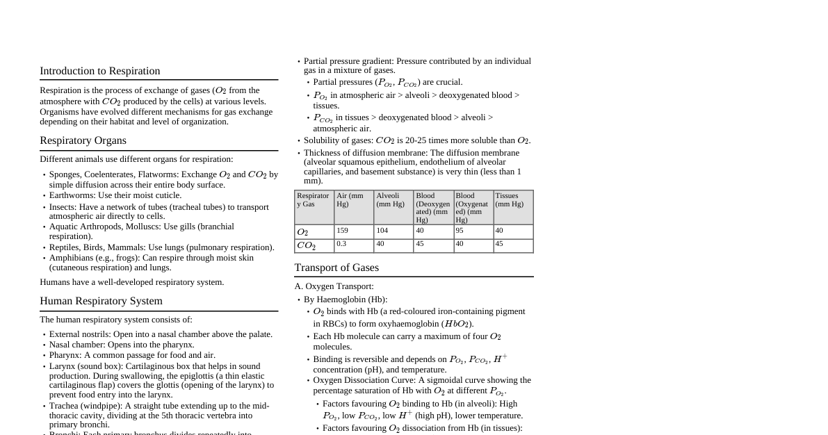
1. Introduction to Respiration Breathing: Physical process of taking in $\text{O}_2$ and giving out $\text{CO}_2$. Respiration: Biochemical process where food is broken down to release energy. Respiratory Organs: Lower invertebrates (sponges, coelenterates, flatworms): Simple diffusion across body surface. Earthworms: Moist cuticle. Insects: Tracheal tubes. Aquatic arthropods/molluscs: Gills (branchial respiration). Terrestrial forms (reptiles, birds, mammals): Lungs (pulmonary respiration). Amphibians (e.g., frogs): Moist skin (cutaneous respiration) and lungs. 2. Human Respiratory System Pathway of Air: External Nostrils Nasal Chamber (via pharynx) Pharynx (common passage for food and air) Larynx (voice box, prevents food entry into trachea via epiglottis) Trachea (windpipe, C-shaped cartilaginous rings) Primary Bronchi (left & right) Secondary Bronchi Tertiary Bronchi Bronchioles (terminal $\rightarrow$ respiratory) Alveoli (site of gas exchange) Lungs: Paired, located in thoracic cavity. Covered by double-layered pleura with pleural fluid (reduces friction). Right lung: 3 lobes; Left lung: 2 lobes. Conducting Part: External nostrils to terminal bronchioles. Transports air, humidifies, warms, filters. Respiratory or Exchange Part: Alveoli and their ducts. Site of actual gas exchange. Thoracic Cavity: Air-tight chamber. Bounded dorsally by vertebral column, ventrally by sternum, laterally by ribs, and inferiorly by diaphragm. 3. Mechanism of Breathing Involves creation of pressure gradients. Inspiration (active process): Diaphragm contracts (flattens), increasing volume of thoracic cavity (antero-posterior axis). External intercostal muscles contract, lifting ribs and sternum (dorso-ventral axis), increasing thoracic volume. Overall increase in thoracic volume $\rightarrow$ decrease in intra-pulmonary pressure (below atmospheric pressure). Air rushes into lungs. Expiration (passive process): Diaphragm relaxes (domes upwards), reducing thoracic volume. External intercostal muscles relax, ribs and sternum return to normal position, reducing thoracic volume. Overall decrease in thoracic volume $\rightarrow$ increase in intra-pulmonary pressure (above atmospheric pressure). Air is expelled from lungs. Forced Expiration: Requires contraction of internal intercostal and abdominal muscles. Normal breathing rate: $12-16$ times/minute. 4. Respiratory Volumes and Capacities Measured by spirometer (except Residual Volume). Tidal Volume (TV): Volume of air inspired/expired during normal respiration. $\approx 500 \text{ mL}$. Inspiratory Reserve Volume (IRV): Additional volume of air inspired by a forceful inspiration. $\approx 2500-3000 \text{ mL}$. Expiratory Reserve Volume (ERV): Additional volume of air expired by a forceful expiration. $\approx 1000-1100 \text{ mL}$. Residual Volume (RV): Volume of air remaining in lungs after a forceful expiration. $\approx 1100-1200 \text{ mL}$. (Cannot be measured by spirometer) Inspiratory Capacity (IC): Total volume of air a person can inspire after a normal expiration. $\text{IC} = \text{TV} + \text{IRV}$. Expiratory Capacity (EC): Total volume of air a person can expire after a normal inspiration. $\text{EC} = \text{TV} + \text{ERV}$. Functional Residual Capacity (FRC): Volume of air remaining in lungs after a normal expiration. $\text{FRC} = \text{ERV} + \text{RV}$. Vital Capacity (VC): Maximum volume of air a person can breathe in after a forced expiration, or maximum volume of air a person can breathe out after a forced inspiration. $\text{VC} = \text{ERV} + \text{TV} + \text{IRV}$. Total Lung Capacity (TLC): Total volume of air accommodated in the lungs at the end of a forced inspiration. $\text{TLC} = \text{RV} + \text{ERV} + \text{TV} + \text{IRV} = \text{VC} + \text{RV}$. 5. Exchange of Gases Occurs at two sites: Alveoli (between air and blood). Tissues (between blood and tissues). Exchange occurs by simple diffusion based on: Partial pressure gradients of $\text{O}_2$ and $\text{CO}_2$. Solubility of gases. Thickness of diffusion membrane. Partial Pressure (p): Pressure contributed by an individual gas in a mixture. Diffusion Membrane: Three major layers: Thin squamous epithelium of alveoli. Endothelium of alveolar capillaries. Basement substance (thin membrane) between them. Total thickness is much less than a millimeter. Partial Pressures (in mm Hg) Gas Atmospheric Air Alveoli Blood (Deoxygenated) Blood (Oxygenated) Tissues $\text{pO}_2$ $159$ $104$ $40$ $95$ $40$ $\text{pCO}_2$ $0.3$ $40$ $45$ $40$ $45$ $\text{CO}_2$ has $20-25$ times higher solubility than $\text{O}_2$. 6. Transport of Gases 6.1. Oxygen Transport $97\%$ transported by Red Blood Cells (RBCs) via hemoglobin. $3\%$ transported in dissolved state in plasma. Hemoglobin (Hb): Red-coloured iron-containing pigment. Each Hb molecule can bind to 4 $\text{O}_2$ molecules. Oxyhemoglobin: $\text{HbO}_2$. Binding is reversible. Oxygen-Dissociation Curve: Sigmoid (S-shaped) curve showing the percentage saturation of hemoglobin with $\text{O}_2$ at various $\text{pO}_2$ values. Factors affecting binding (shift curve to right/left): $\text{pCO}_2$ $\text{H}^+$ concentration (pH) Temperature $2,3$-BPG In alveoli: High $\text{pO}_2$, low $\text{pCO}_2$, less $\text{H}^+$ (higher pH), lower temperature $\rightarrow$ favors $\text{O}_2$ binding to Hb. In tissues: Low $\text{pO}_2$, high $\text{pCO}_2$, high $\text{H}^+$ (lower pH), higher temperature $\rightarrow$ favors $\text{O}_2$ dissociation from Hb (Bohr effect). 6.2. Carbon Dioxide Transport $7\%$ transported in dissolved state in plasma. $20-25\%$ transported by Hb as Carbaminohemoglobin ($\text{HbCO}_2$). Binding is related to $\text{pCO}_2$ and $\text{pO}_2$. In tissues: High $\text{pCO}_2$, low $\text{pO}_2 \rightarrow$ favors $\text{CO}_2$ binding to Hb. In alveoli: Low $\text{pCO}_2$, high $\text{pO}_2 \rightarrow$ favors dissociation of $\text{CO}_2$ from Hb (Haldane effect). $70\%$ transported as Bicarbonate Ions ($\text{HCO}_3^-$). In RBCs, $\text{CO}_2 + \text{H}_2\text{O} \xrightarrow{\text{Carbonic Anhydrase}} \text{H}_2\text{CO}_3 \xrightarrow{} \text{H}^+ + \text{HCO}_3^-$. $\text{HCO}_3^-$ moves into plasma, $\text{Cl}^-$ moves into RBCs (Chloride shift or Hamburger phenomenon). In alveoli, the reaction reverses, $\text{CO}_2$ is released. 7. Regulation of Respiration Mainly regulated by the nervous system. Respiratory Rhythm Centre: Medulla oblongata (primary control). Pneumotaxic Centre: Pons Varolii. Can moderate the functions of the rhythm centre. Reduces duration of inspiration, thus altering respiratory rate. Chemosensitive Area: Adjacent to rhythm centre, highly sensitive to $\text{CO}_2$ and $\text{H}^+$ ions. Increase in $\text{CO}_2$ or $\text{H}^+$ stimulates this centre, signaling the rhythm centre to increase respiratory rate to eliminate $\text{CO}_2$. Receptors associated with aortic arch and carotid artery also detect changes in $\text{CO}_2$ and $\text{H}^+$ concentrations and send signals to rhythm centre. Role of $\text{O}_2$ in respiratory regulation is quite insignificant. 8. Disorders of Respiratory System Asthma: Difficulty in breathing due to inflammation of bronchi and bronchioles. Emphysema: Chronic disorder where alveolar walls are damaged, decreasing respiratory surface. Major cause is cigarette smoking. Occupational Respiratory Disorders: Caused by prolonged exposure to dust (e.g., grinding, stone-breaking). Leads to inflammation and fibrosis (proliferation of fibrous tissue). e.g., Silicosis, Asbestosis. Protective masks are recommended.