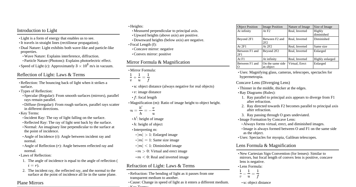
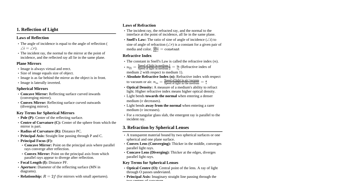
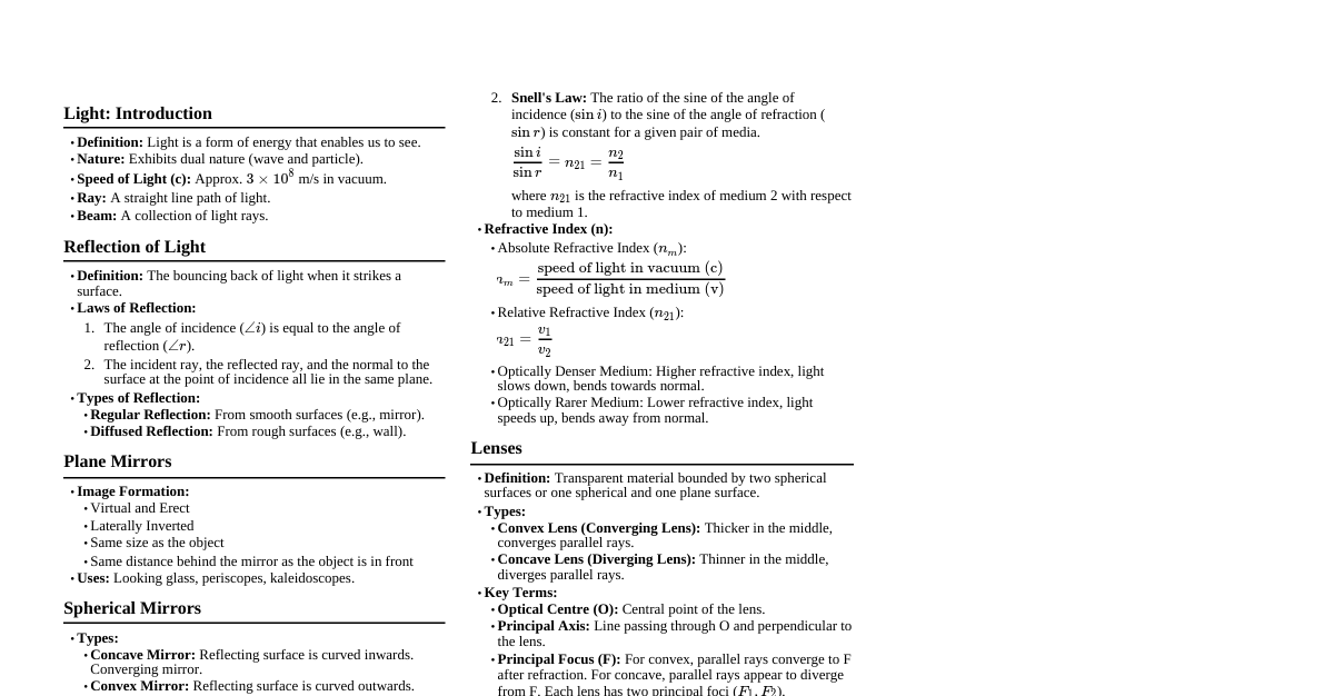
Vision & Refraction Tools
Cheatsheet Content
### Color Vision Theories Color vision is the ability of the eye and brain to distinguish objects based on the wavelengths of light they reflect or emit. #### Trichromatic Theory (Young-Helmholtz) - **Concept:** Proposes that the human eye has three types of cone photoreceptors, each sensitive to different wavelengths of light: - **Short (S) cones:** Most sensitive to blue light (approx. 420-440 nm). - **Medium (M) cones:** Most sensitive to green light (approx. 530-540 nm). - **Long (L) cones:** Most sensitive to red-yellow light (approx. 560-580 nm). - **Mechanism:** All colors are perceived by the brain through the differential stimulation of these three cone types. For example, yellow light stimulates both L and M cones, while purple light stimulates S and L cones. - **Evidence:** Explains forms of color blindness (e.g., protanopia, deuteranopia, tritanopia) where one or more cone types are deficient or absent. #### Opponent-Process Theory (Hering) - **Concept:** Suggests that color perception is controlled by three opponent systems in the visual pathway, where opposing colors cannot be perceived simultaneously: - **Red-Green system:** Processes red vs. green signals. - **Blue-Yellow system:** Processes blue vs. yellow signals. - **Black-White system:** Processes light vs. dark signals (luminance). - **Mechanism:** When one color in an opponent pair is stimulated, the other is inhibited. For example, staring at a red object for a long time can lead to a green afterimage when looking at a white surface, due to fatigue of the red-sensitive cells and subsequent activation of green-sensitive cells. - **Evidence:** Explains phenomena like complementary afterimages and the impossibility of perceiving "reddish-green" or "yellowish-blue." It also accounts for color vision beyond the retina, at the ganglion cell and cortical levels. - **Integration:** Modern understanding combines both theories: trichromacy operates at the photoreceptor level, while opponent processes occur in later stages of visual processing (ganglion cells, LGN, visual cortex). ### Color Arrangement Test (Farnsworth D-15 / 100-Hue) These tests assess an individual's ability to discriminate between subtle differences in color hues. #### Farnsworth D-15 (Dichotomous 15-Hue Test) - **Purpose:** Screens for moderate to severe color vision defects (dichromacy or severe anomalous trichromacy). Not sensitive enough for mild defects. - **Procedure:** The patient is presented with 15 caps of varying hues and a fixed reference cap. They must arrange the 15 caps in a continuous color sequence, starting from the reference cap. - **Interpretation:** - **Normal Vision:** The caps are arranged in a perfect circle or a smooth elliptical shape on a scoring sheet. - **Defective Vision:** Errors in arrangement indicate a specific axis of confusion, suggesting the type of color vision defect (e.g., protan, deutan, tritan). Lines crossing the center of the scoring circle indicate an axis of confusion. - **Scoring:** Errors are plotted on a polar diagram. Lines connecting incorrectly placed caps reveal the axis of confusion. - **Clinical Significance:** Identifies individuals who may have difficulty with tasks requiring good color discrimination, such as certain professions (e.g., electricians, pilots, graphic designers). #### Farnsworth-Munsell 100-Hue Test - **Purpose:** A more detailed and sensitive test used to classify and quantify the degree of color vision deficiency (CVD), including mild defects, and to detect acquired color vision abnormalities. - **Procedure:** Consists of four trays, each containing 21-22 caps of varying hues. The patient arranges the caps in order of hue from a fixed reference cap at each end of the tray. - **Interpretation:** - **Normal Vision:** Low total error score, with errors distributed evenly. - **Defective Vision:** Higher total error score. The pattern of errors on a polar diagram indicates the type of defect (protan, deutan, tritan) and its severity. Acquired defects often show a tritan axis. - **Total Error Score (TES):** Calculated by summing the differences in cap numbers for each misplaced cap. Lower scores indicate better color discrimination. - **Clinical Significance:** Used for research, occupational screening where precise color discrimination is critical, and monitoring changes in color vision due to disease (e.g., optic nerve disease, macular degeneration). ### Autokeratometer An autokeratometer is an automated device used to measure the curvature of the anterior surface of the cornea. - **Principle:** It projects a series of infrared light rings (mires) onto the cornea. The cornea acts as a convex mirror, reflecting these mires. The instrument then analyzes the size and shape of the reflected mires. - **Measurement:** It primarily measures the radius of curvature in two principal meridians (usually the steepest and flattest) and the angle between them. From these measurements, it calculates: - **Corneal curvature:** Expressed in millimeters of radius. - **Corneal power:** Expressed in diopters (D), using a standard refractive index for the cornea (typically 1.3375). - **Corneal astigmatism:** The difference in power between the two principal meridians, and the axis of the astigmatism. - **Procedure:** The patient places their chin on a rest and forehead against a support. They focus on an internal fixation target. The operator aligns the instrument, and the measurement is taken automatically, often in a few seconds. - **Advantages:** - **Speed and Ease of Use:** Rapid measurements, minimal operator skill required. - **Objectivity:** Reduces subjective bias inherent in manual keratometry. - **Repeatability:** Good for monitoring changes in corneal curvature (e.g., after contact lens wear, refractive surgery). - **Disadvantages:** - **Limited Scope:** Measures only the central 3-4 mm of the cornea. - **Accuracy:** Can be affected by tear film abnormalities, small pupils, or patient fixation issues. - **Peripheral Cornea:** Does not provide information on peripheral corneal shape, which is crucial for certain contact lens fittings or conditions like keratoconus. - **Clinical Significance:** - **Contact Lens Fitting:** Essential for determining base curve and astigmatic parameters. - **Refractive Surgery (LASIK, PRK):** Pre-operative assessment and post-operative monitoring of corneal changes. - **Intraocular Lens (IOL) Calculation:** Contributes to the calculation of IOL power for cataract surgery. - **Diagnosis of Corneal Conditions:** Helps detect and monitor conditions like keratoconus (though topography is superior for this). ### Autorefractometer An autorefractometer is an objective, automated device used to measure the refractive error of the eye. - **Principle:** It projects infrared light into the eye and measures the light reflected from the retina. The instrument then analyzes how the light has changed as it passed through the ocular media and back, determining the lens power needed to bring the light into sharp focus on the retina. - **Measurement:** It typically provides a reading for: - **Sphere (Sph):** Myopia (negative power) or hyperopia (positive power). - **Cylinder (Cyl):** Amount of astigmatism. - **Axis (Ax):** Orientation of the astigmatism. - **Pupil Distance (PD):** Often measured automatically. - **Procedure:** Similar to an autokeratometer, the patient places their chin and forehead on supports and fixates on an internal target (often an image that appears to recede and advance to relax accommodation). The instrument takes multiple readings automatically. - **Advantages:** - **Speed and Efficiency:** Quick and easy to operate, ideal for screening many patients. - **Objectivity:** Does not require subjective responses from the patient, useful for children, uncooperative patients, or those with language barriers. - **Baseline Data:** Provides a good starting point for subjective refraction. - **Disadvantages:** - **Accommodation:** Can sometimes induce accommodation, leading to a more myopic reading, especially in younger patients. Some devices incorporate "fogging" techniques to mitigate this. - **Accuracy:** Less precise than subjective refraction and requires an experienced clinician to refine the prescription. - **Ocular Media Opacities:** Cataracts or other media opacities can interfere with accurate readings. - **Clinical Significance:** - **Refraction Baseline:** Provides an initial estimate of the patient's refractive error, saving time during subjective refraction. - **Screening:** Widely used in general eye exams, school screenings, and occupational health. - **Paediatric Optometry:** Very useful for young children who cannot reliably respond during subjective refraction. - **Monitoring:** Helps track refractive changes over time. ### Aberrometer An aberrometer is a sophisticated device that measures higher-order aberrations (HOAs) of the eye, which are more complex optical errors than sphere and cylinder. - **Principle:** Most modern aberrometers use the **Hartmann-Shack wavefront sensor** method. 1. A low-power infrared light beam is projected into the eye, creating a point source image on the retina. 2. The light reflected from the retina passes back out through the ocular media. 3. Instead of being a perfect plane (as it would be in a perfectly corrected eye), the wavefront emerging from an aberrated eye is distorted. 4. This distorted wavefront passes through a lenslet array (a grid of tiny lenses) on the aberrometer. 5. Each lenslet focuses a portion of the wavefront onto a CCD camera. 6. The displacement of these focal spots from their ideal positions (if the eye were perfect) is measured. 7. Mathematical algorithms (Zernike polynomials) are used to reconstruct the shape of the wavefront and quantify the various aberrations. - **Measurement:** Provides a detailed map of the eye's optical imperfections, including: - **Lower-Order Aberrations (LOAs):** Sphere (defocus), cylinder (astigmatism), and prism. These account for about 90% of a typical eye's aberrations. - **Higher-Order Aberrations (HOAs):** Coma, trefoil, spherical aberration, etc. These can significantly impact vision quality, especially in low light or with large pupils, causing symptoms like glare, halos, starbursts, and reduced contrast sensitivity. - **Procedure:** Similar to autorefractometry, the patient fixates on a target. The device takes a rapid measurement, providing a comprehensive wavefront analysis. - **Advantages:** - **Comprehensive Optical Analysis:** Measures both LOAs and HOAs, providing a complete picture of the eye's optics. - **Improved Visual Quality:** Allows for "wavefront-guided" corrections (e.g., custom LASIK) that can provide vision potentially superior to traditional spectacle or contact lens correction. - **Objective and Precise:** Highly accurate and repeatable measurements. - **Detection of Subtle Anomalies:** Can detect optical irregularities not found by standard refraction. - **Disadvantages:** - **Pupil Size Dependency:** HOA measurements are highly dependent on pupil size. - **Cost and Complexity:** More expensive and complex than autorefractometers. - **Dynamic Eye:** The eye's aberrations can change with accommodation, tear film stability, and pupil size. - **Clinical Significance:** - **Refractive Surgery (Custom LASIK/PRK):** Essential for wavefront-guided treatments, aiming to correct HOAs and improve quality of vision. - **Complex Contact Lens Fitting:** Can aid in designing custom contact lenses for irregular corneas (e.g., keratoconus). - **Diagnosis of Ocular Pathology:** Can help identify optical changes associated with corneal diseases, cataracts, or intraocular lens decentration. - **Research:** Used extensively in optics and visual science research.