ICSE Biology Diagrams
Cheatsheet Content
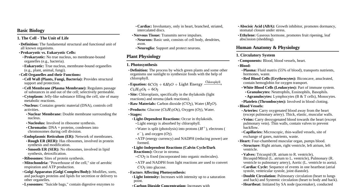
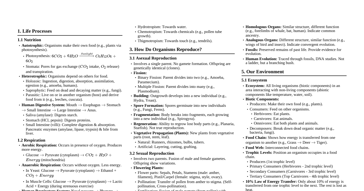
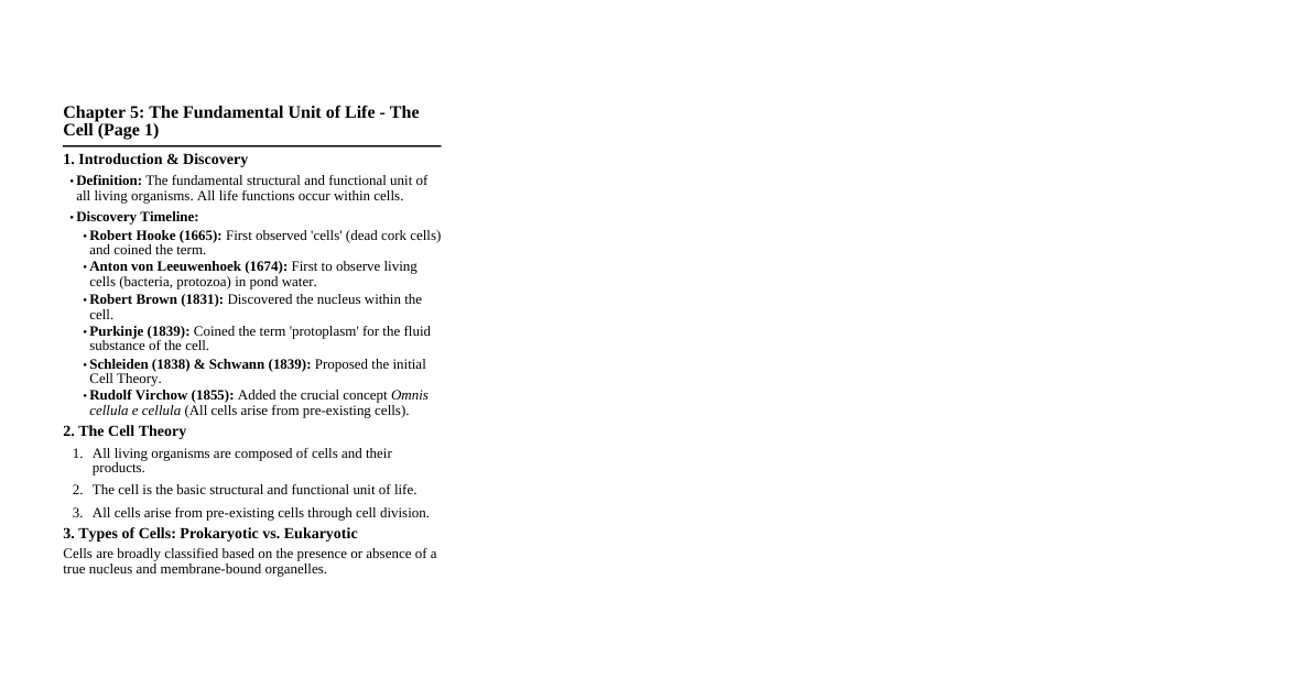
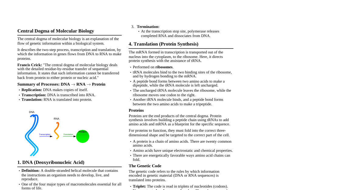
Cell Structure and Division Animal Cell: A typical animal cell showing: Cell membrane. Cytoplasm. Nucleus (with nuclear membrane, nucleolus, chromatin material). Mitochondria. Endoplasmic Reticulum (rough and smooth). Ribosomes. Golgi apparatus. Centrosome (with centrioles). Lysosomes, vacuoles (if any). Plant Cell: A typical plant cell showing: Cell wall. Cell membrane. Cytoplasm. Nucleus (with nuclear membrane, nucleolus, chromatin material). Mitochondria. Chloroplasts. Large central vacuole. Endoplasmic Reticulum, Ribosomes, Golgi apparatus. Mitosis: Key stages of mitosis in an animal or plant cell: Prophase: Chromatin condenses, spindle fibers form. Metaphase: Chromosomes align at the metaphase plate. Anaphase: Sister chromatids separate and move to opposite poles. Telophase: Chromosomes decondense, nuclear envelopes reform, cytokinesis begins. Photosynthesis and Transpiration Chloroplast: Diagram showing the internal structure of a chloroplast: Outer and inner membranes. Stroma. Grana (stacks of thylakoids). Thylakoids. Stomata: Diagrams of both open and closed stomata, showing: Guard Cells: Bean-shaped cells, regulate stomatal pore. Stomatal Pore: Opening for gas exchange. Subsidiary Cells: Accessory cells surrounding guard cells. Chloroplasts within guard cells. Transpiration Experiments: Bell Jar Experiment: Potted plant covered with a bell jar, showing water droplets on the inner surface. Potometer: Apparatus to measure the rate of water uptake (and thus transpiration) by a leafy shoot. Circulatory System Human Heart: Internal structure showing: Four chambers: Right Atrium, Left Atrium, Right Ventricle, Left Ventricle. Major blood vessels: Aorta, Pulmonary Artery, Pulmonary Veins, Superior Vena Cava, Inferior Vena Cava. Valves: Tricuspid valve, Bicuspid (Mitral) valve, Pulmonary valve, Aortic valve. Septum. Blood Vessels: Transverse sections comparing: Artery: Thick, muscular, elastic walls, narrow lumen. Vein: Thinner walls, wider lumen, presence of valves. Capillary: Extremely thin (one cell thick) walls. Double Circulation: Schematic diagram illustrating: Pulmonary circulation (heart to lungs and back). Systemic circulation (heart to body and back). Excretory System Human Excretory System: Labeling of major organs: Kidneys (paired). Ureters (tubes from kidneys to bladder). Urinary Bladder. Urethra. Nephron: Detailed structure of the functional unit of the kidney: Bowman's Capsule: Cup-shaped structure. Glomerulus: Capillary network inside Bowman's capsule. Renal Tubule: Proximal Convoluted Tubule (PCT). Loop of Henle (descending and ascending limbs). Distal Convoluted Tubule (DCT). Collecting Duct. Associated blood vessels (afferent/efferent arterioles, peritubular capillaries). Nervous System and Sense Organs Neuron (Nerve Cell): Structure showing: Cell Body (Soma): Contains nucleus. Dendrites: Receive signals. Axon: Transmits signals away from cell body. Myelin Sheath: Insulating layer. Nodes of Ranvier: Gaps in myelin sheath. Synaptic Knobs/Terminals: Endings that transmit signals to other cells. Reflex Arc: Diagram showing the pathway of a reflex action: Stimulus and Receptor . Sensory Neuron (afferent). Relay Neuron (Interneuron) in spinal cord. Motor Neuron (efferent). Effector (muscle or gland) and Response . Human Eye: Longitudinal section showing: Cornea: Transparent outer layer. Iris: Controls pupil size. Pupil: Opening in the center of the iris. Lens: Focuses light. Retina: Light-sensitive layer with photoreceptors (rods and cones). Optic Nerve: Transmits visual information to brain. Ciliary Body, Suspensory Ligaments, Vitreous Humor, Aqueous Humor, Blind Spot, Yellow Spot/Macula. Human Ear: Diagram showing the three main parts: Outer Ear: Pinna (auricle), Ear canal (auditory meatus). Middle Ear: Eardrum (tympanic membrane), Ossicles (malleus, incus, stapes), Eustachian tube. Inner Ear: Cochlea (hearing), Semicircular Canals (balance), Auditory nerve. Endocrine System Human Endocrine Glands: Location of major glands in the body: Pituitary Gland (base of brain). Thyroid Gland (neck). Pancreas (abdomen). Adrenal Glands (on top of kidneys). Testes (males) / Ovaries (females). Reproductive System Human Reproductive System (Male): Diagram showing: Testis, Epididymis, Vas deferens, Seminal vesicles, Prostate gland, Urethra, Penis. Human Reproductive System (Female): Diagram showing: Ovaries, Fallopian tubes (oviducts), Uterus, Cervix, Vagina. Fertilization and Implantation: Schematic representation from ovulation to implantation of the embryo in the uterine wall. Human Respiration Human Respiratory System: Labeling of pathways and organs: Nasal passage, Pharynx, Larynx (voice box). Trachea (windpipe) with cartilaginous rings. Bronchi, Bronchioles, Lungs (right and left). Diaphragm. Detailed structure of Alveoli: Showing thin walls, blood capillaries, gas exchange. Mechanism of Breathing: Diagrams illustrating inhalation and exhalation, showing: Movement of diaphragm. Movement of rib cage. Volume changes in thoracic cavity. Air pressure changes. Absorption by Roots Root Hair Cell: A specialized epidermal cell for water and mineral absorption, showing: Elongated root hair extension. Cell wall. Cell membrane. Cytoplasm. Nucleus. Large central vacuole. Osmosis Experiment: Setup using a thistle funnel or potato osmometer to demonstrate osmosis.