Plant Anatomy (NEET Advanced)
Cheatsheet Content
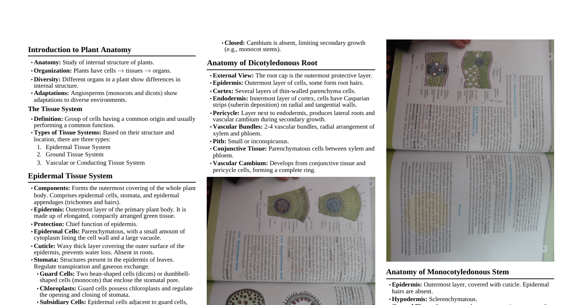
Plant Anatomy: Basic Concepts Anatomy: Study of internal structure of plants. Plants are organized into cells $\rightarrow$ tissues $\rightarrow$ organs. Monocots and dicots show anatomical differences and adaptations to diverse environments. 1. Tissue Systems Based on structure and location, there are three types: 1.1. Epidermal Tissue System Outer-most covering: Comprises epidermal cells, stomata, and epidermal appendages (trichomes and hairs). Epidermis: Outermost layer of primary plant body, usually single-layered. Made of elongated, compactly arranged parenchymatous cells with small cytoplasm and large vacuole. Cuticle: Waxy thick layer on outer epidermis (absent in roots); prevents water loss. Stomata: Structures in leaf epidermis for transpiration and gaseous exchange. Composed of two guard cells enclosing a stomatal pore. Bean-shaped in dicots, dumb-bell shaped in grasses. Outer walls of guard cells are thin, inner walls are highly thickened. Guard cells possess chloroplasts and regulate stomatal opening/closing. Subsidiary cells: Specialized epidermal cells near guard cells. Stomatal Apparatus: Stomatal aperture + guard cells + surrounding subsidiary cells. Epidermal Appendages: Root Hairs: Unicellular elongations of epidermal cells; absorb water and minerals. Trichomes: Epidermal hairs on stem (shoot system); usually multicellular, branched/unbranched, soft/stiff, or secretory; prevent water loss. 1.2. Ground Tissue System All tissues except epidermis and vascular bundles. Consists of simple tissues: parenchyma, collenchyma, and sclerenchyma. Parenchymatous cells: Present in cortex, pericycle, pith, medullary rays (primary stems/roots). Mesophyll: Ground tissue in leaves, consists of thin-walled chloroplast-containing cells. 1.3. Vascular Tissue System Complex tissues: xylem and phloem, together form vascular bundles . Vascular Bundles Types: Open: Cambium present between xylem and phloem (e.g., dicot stems); can form secondary xylem and phloem (secondary growth). Closed: No cambium (e.g., monocot stems); no secondary growth. Radial: Xylem and phloem arranged alternately on different radii (e.g., roots). Conjoint: Xylem and phloem on the same radius (e.g., stems and leaves). Phloem usually located on outer side of xylem. 2. Anatomy of Dicotyledonous and Monocotyledonous Plants 2.1. Dicotyledonous Root (e.g., Sunflower) Epiblema: Outermost layer, cells protrude as unicellular root hairs. Cortex: Several layers of thin-walled parenchyma cells with intercellular spaces. Endodermis: Innermost layer of cortex, single layer of barrel-shaped cells, no intercellular spaces. Tangential and radial walls have deposition of water-impermeable, waxy suberin called casparian strips . Pericycle: Few layers of thick-walled parenchymatous cells next to endodermis. Initiation of lateral roots and vascular cambium during secondary growth occurs here. Pith: Small or inconspicuous. Conjunctive Tissue: Parenchymatous cells between xylem and phloem. Usually 2 to 4 xylem and phloem patches. Cambium ring develops later between xylem and phloem. Stele: All tissues inside endodermis (pericycle, vascular bundles, pith). 2.2. Monocotyledonous Root Similar to dicot root in many respects (epidermis, cortex, endodermis, pericycle, vascular bundles, pith). Xylem Bundles: Usually more than six (polyarch condition), unlike dicot roots with fewer bundles. Pith: Large and well-developed. No secondary growth. 2.3. Dicotyledonous Stem Epidermis: Outermost protective layer, covered with thin cuticle, may bear trichomes and few stomata. Cortex: Multiple layers between epidermis and pericycle, divided into three sub-zones: Hypodermis: Few layers of collenchymatous cells below epidermis; provides mechanical strength. Cortical layers: Rounded, thin-walled parenchymatous cells with conspicuous intercellular spaces. Endodermis: Innermost layer of cortex, rich in starch grains (also called starch sheath ). Pericycle: Present on inner side of endodermis and above phloem, in semi-lunar patches of sclerenchyma. Medullary rays: Few layers of radially placed parenchymatous cells between vascular bundles. Vascular Bundles: Arranged in a ring (characteristic of dicot stem); conjoint, open, with endarch protoxylem. Pith: Large number of rounded, parenchymatous cells with large intercellular spaces in central portion. 2.4. Monocotyledonous Stem Hypodermis: Sclerenchymatous. Vascular Bundles: Scattered, conjoint, closed, each surrounded by a sclerenchymatous bundle sheath. Peripheral bundles generally smaller than centrally located ones. Phloem parenchyma is absent. Water-containing cavities present within vascular bundles. Ground Tissue: Large, conspicuous parenchymatous ground tissue. 2.5. Dorsiventral (Dicotyledonous) Leaf Epidermis: Covers both upper (adaxial) and lower (abaxial) surfaces, conspicuous cuticle. Abaxial epidermis usually bears more stomata; adaxial epidermis may lack stomata. Mesophyll: Tissue between upper and lower epidermis; contains chloroplasts, performs photosynthesis. Made of two types of cells: Palisade parenchyma: Adaxially placed, elongated cells arranged vertically and parallel. Spongy parenchyma: Below palisade cells, oval/round, loosely arranged, numerous large spaces and air cavities. Vascular System: Vascular bundles seen in veins and midrib. Size depends on vein size. Vascular bundles surrounded by thick-walled bundle sheath cells . 2.6. Isobilateral (Monocotyledonous) Leaf Similar to dorsiventral leaf but with key differences: Stomata present on both adaxial and abaxial epidermis. Mesophyll not differentiated into palisade and spongy parenchyma. In grasses, certain adaxial epidermal cells along veins modify into large, empty, colorless bulliform cells . When turgid, leaf surface exposed. When flaccid (due to water stress), leaves curl inwards to minimize water loss. Parallel venation reflected in near similar sizes of vascular bundles (except main veins).