Structural Org. in Animals (NEET)
Cheatsheet Content
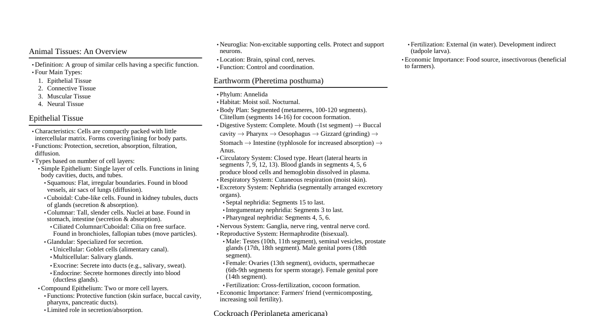
I. Tissues Group of similar cells along with intercellular material performing a specific function. Four types: Epithelial, Connective, Muscular, Neural. II. Epithelial Tissue Covers body surface, lines body cavities, ducts, and forms glands. Cells are tightly packed with little intercellular matrix. Types: Simple Epithelium: Single layer of cells. Squamous: Flat, irregular boundaries. Found in blood vessels, air sacs of lungs. Forms diffusion boundary. Cuboidal: Cube-like cells. Found in ducts of glands, tubular parts of nephrons. Secretion and absorption. Columnar: Tall, slender cells, nuclei at base. Found in stomach, intestine. Secretion and absorption. If they bear cilia: Ciliated epithelium (bronchioles, fallopian tubes) - move particles/mucus in a specific direction. Glandular: Specialised for secretion. Unicellular: Isolated glandular cells (Goblet cells of alimentary canal). Multicellular: Cluster of cells (Salivary gland). Exocrine: Secrete products through ducts (mucus, saliva, oil, milk, digestive enzymes). Endocrine: Ductless glands, secrete hormones directly into fluid bathing gland. Compound Epithelium: Two or more cell layers. Protection against chemical and mechanical stresses. Limited role in secretion and absorption. Found in skin, buccal cavity, pharynx, inner lining of salivary gland ducts, pancreatic ducts. Cell Junctions: Tight Junctions: Stop substances from leaking across tissue. Adhering Junctions: Cement neighbouring cells together. Gap Junctions: Facilitate communication between cells by connecting cytoplasm, rapid transfer of ions, small molecules, large molecules. III. Connective Tissue Most abundant and widely distributed tissue. Link and support other tissues/organ. Cells are widely spaced, embedded in abundant intercellular matrix (non-living ground substance + fibers). Fibers: Collagen (strength), Elastin (elasticity), Reticular (support). Cells: Fibroblasts (produce/secrete fibers), Macrophages (phagocytic), Mast cells (secrete histamine, serotonin, heparin). Types: Loose Connective Tissue: Areolar: Beneath skin. Support framework for epithelium, contains fibroblasts, macrophages, mast cells. Adipose: Beneath skin. Stores fat, acts as insulator, fat storage. Dense Connective Tissue: Fibers and fibroblasts compactly packed. Dense Regular: Collagen fibers in parallel rows. Tendons: Connect muscle to bone. Ligaments: Connect bone to bone. Dense Irregular: Fibers oriented differently. Found in skin. Specialised Connective Tissue: Cartilage: Solid, pliable, resists compression. Chondrocytes enclosed in small cavities within matrix. Found in nose tip, outer ear, joints, vertebral column. Bone: Hard, non-pliable, rich in calcium salts and collagen. Osteocytes in lacunae. Provides structural frame, supports, protects organs, muscle attachment. Blood: Fluid connective tissue. Plasma (fluid matrix), RBCs, WBCs, platelets. Transport. IV. Muscular Tissue Specialised for movement. Made of elongated cells called muscle fibers. Fibers contain numerous fine fibrils called myofibrils. Contract in response to stimulation, then relax. Types: Skeletal (Striated/Voluntary): Closely attached to bones. Striations present. Voluntary control. Smooth (Non-striated/Involuntary): Walls of internal organs (stomach, intestine, blood vessels). No striations. Involuntary control. Cardiac (Heart Muscle): Found only in the heart. Striated, involuntary. Cells are branched, interconnections via intercalated discs (gap junctions). V. Neural Tissue Controls body's responsiveness to changing conditions. Composed of neurons (excitatory cells) and neuroglia (support cells). Neurons: Functional unit. Cell body, dendrites, axon. Stimulated, generate and transmit electrical impulses. Neuroglia: Protect and support neurons. Make up more than half the volume of neural tissue. VI. Cockroach (Periplaneta americana) Phylum: Arthropoda, Class: Insecta. Nocturnal, omnivorous, found in damp places. Body Segmentation: Head, Thorax, Abdomen. Exoskeleton: Chitinous, sclerites (tergites dorsally, sternites ventrally) joined by arthrodial membrane. Head: Triangular, anterior, right angles to body axis. Pair of antennae (sensory). Compound eyes. Mouthparts: Labrum (upper lip), mandibles, maxillae, labium (lower lip), hypopharynx (tongue). Thorax: Prothorax, Mesothorax, Metathorax. Each segment bears a pair of walking legs. Forewings (tegmina) from mesothorax (opaque, leathery, cover hindwings). Hindwings from metathorax (transparent, membranous, used in flight). Abdomen: 10 segments. Males: Anal cerci (paired), anal style (paired, short, thread-like, present only in males). Females: Brood or genital pouch (7th, 8th, 9th sterna). Digestive System: Alimentary Canal: Foregut (mouth to gizzard), Midgut (hepatic caeca), Hindgut (ileum, colon, rectum, anus). Gizzard (Proventriculus): Grinding food. Hepatic Caeca: 6-8 blind tubules at junction of foregut and midgut, secrete digestive juice. Malpighian Tubules: 100-150 yellow tubules at junction of midgut and hindgut, excretory. Circulatory System: Open type. Haemocoel (body cavity full of haemolymph). Heart: Elongated, muscular tube in thorax and abdomen, 13 funnel-shaped chambers. Haemolymph: Colourless plasma + haemocytes. Respiratory System: Network of tracheae. Tracheal tubes open via 10 pairs of spiracles (2 thoracic, 8 abdominal). Oxygen directly to cells. Excretory System: Malpighian tubules. Uricotelic (excretes uric acid). Fat body, nephrocytes, uricose glands also help. Nervous System: Segmentally arranged ganglia joined by paired longitudinal connectives on ventral side. Brain (supra-oesophageal ganglion) in head. Most of nervous system in abdomen and thorax. Reproductive System: Dioecious. Male: Pair of testes (4th-6th abdominal segments). Vas deferens, ejaculatory duct, male gonopore. Mushroom gland (6th-7th segments). Phallic glands, external genitalia (male gonapophysis/phallomere). Spermatophores (sperm packets). Female: Pair of ovaries (2-6th abdominal segments). Oviducts, vagina, genital chamber. Spermatheca (stores sperm). Collateral glands. Ootheca (dark reddish to blackish brown capsule containing 14-16 eggs). Development: Paurometabolous (gradual metamorphosis) - nymph to adult. VII. Frog (Rana tigrina) Phylum: Chordata, Class: Amphibia. Can live on land and in fresh water. Poikilotherms (cold-blooded). Camouflage (mimicry) for protection. Aestivation (summer sleep) and hibernation (winter sleep). Body: Head and Trunk. No neck or tail. Skin: Moist, smooth, slippery. Mucus secretion. Always moist. Respiration through skin. Limbs: Forelimbs (4 digits), Hindlimbs (5 digits, webbed - swimming). Sexual Dimorphism: Males have vocal sacs and copulatory pads on first digit of forelimbs. Digestive System: Alimentary canal short (carnivorous). Mouth, pharynx, oesophagus, stomach, intestine, rectum, cloaca. Liver (bile secretion), Pancreas (digestive enzymes). Respiratory System: Air $\to$ nostrils $\to$ buccal cavity $\to$ lungs $\to$ respiration. Cutaneous: Through moist skin (on land and in water). Pulmonary: Through lungs (on land). Buccopharyngeal: Through buccopharyngeal cavity (on land). Circulatory System: Closed type. Well-developed lymphatic system. Heart: 3-chambered (2 atria, 1 ventricle). Sinus venosus: Triangular structure, joins right atrium, receives blood through major veins (venae cavae). Ventricle opens on the ventral side into a sac-like conus arteriosus . RBCs are nucleated. Venous system (hepatic portal system, renal portal system). Excretory System: Pair of kidneys (dark red, bean-like). Ureters, cloaca. Thin-walled urinary bladder is present ventral to the rectum and opens into the cloaca. Rectum also opens into the cloaca. Ureotelic (excretes urea). Presence of finger-like fat bodies attached to kidneys. Nervous System: Central (brain, spinal cord), Peripheral (cranial, spinal nerves), Autonomic. 10 pairs of cranial nerves. Brain: Forebrain: Olfactory lobes, paired cerebral hemispheres, unpaired diencephalon. Midbrain: Pair of optic lobes. Hindbrain: Cerebellum and medulla oblongata. Medulla oblongata passes through foramen magnum and continues as spinal cord. Sense Organs: Eyes and ears are well organized. Other sense organs are cellular aggregations around nerve endings. Eyes (simple, protected by nictitating membrane), tympanum (ear), olfactory lobes, taste buds, touch receptors. Reproductive System: Dioecious. Male: Pair of testes (yellowish, ovoid, attached to kidneys). Peritoneum called mesorchium attaches testes to kidney. Vasa efferentia (10-12), kidneys $\to$ Bidder’s canal $\to$ ureter (urinogenital duct). Cloaca. Female: Pair of ovaries (near kidneys). Oviducts, cloaca. Female can lay 2500–3000 ova at a time. Fertilisation external, in water. Development indirect (larval stage: tadpole).