Anatomy of Flowering Plants
Cheatsheet Content
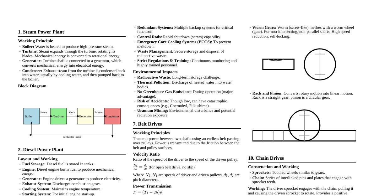
### Introduction to Plant Anatomy - **Anatomy:** Study of internal structure of plants. - **Organization:** Plants have cells $\rightarrow$ tissues $\rightarrow$ organs. - **Diversity:** Different organs in a plant show differences in internal structure. - **Adaptations:** Angiosperms (monocots and dicots) show adaptations to diverse environments. #### The Tissue System - **Definition:** Group of cells having a common origin and usually performing a common function. - **Types of Tissue Systems:** Based on their structure and location, there are three types: 1. Epidermal Tissue System 2. Ground Tissue System 3. Vascular or Conducting Tissue System ### Epidermal Tissue System - **Components:** Forms the outermost covering of the whole plant body. Comprises epidermal cells, stomata, and epidermal appendages (trichomes and hairs). - **Epidermis:** Outermost layer of the primary plant body. It is made up of elongated, compactly arranged green tissue. - **Protection:** Chief function of epidermis. - **Epidermal Cells:** Parenchymatous, with a small amount of cytoplasm lining the cell wall and a large vacuole. - **Cuticle:** Waxy thick layer covering the outer surface of the epidermis, prevents water loss. Absent in roots. - **Stomata:** Structures present in the epidermis of leaves. Regulate transpiration and gaseous exchange. - **Guard Cells:** Two bean-shaped cells (dicots) or dumbbell-shaped cells (monocots) that enclose the stomatal pore. - **Chloroplasts:** Guard cells possess chloroplasts and regulate the opening and closing of stomata. - **Subsidiary Cells:** Epidermal cells adjacent to guard cells, sometimes specialized in shape and size. - **Stomatal Apparatus:** Stomatal pore, guard cells, and surrounding subsidiary cells. - **Epidermal Appendages:** - **Epidermal Hairs:** Unicellular or multicellular extensions of epidermal cells on stems. - **Trichomes:** Root hairs are unicellular elongations of the epidermal cells, which absorb water and minerals from the soil. On the stem, epidermal hairs are called trichomes. Trichomes can be branched or unbranched, soft or stiff, and may even be secretory. They help prevent water loss due to transpiration. ### Ground Tissue System - **Components:** All tissues except epidermis and vascular bundles constitute the ground tissue. - **Types of Ground Tissues:** Consists of simple tissues such as parenchyma, collenchyma, and sclerenchyma. - **Location:** - **Parenchymatous cells:** Usually present in cortex, pericycle, pith, and medullary rays in primary stems and roots. - **Leaves:** Ground tissue consists of thin-walled chloroplast-containing cells called mesophyll. ### Vascular Tissue System - **Components:** Consists of complex tissues (xylem and phloem). - **Vascular Bundles:** Xylem and phloem together form vascular bundles. - **Arrangement of Vascular Bundles:** - **Radial:** Xylem and phloem are arranged in an alternate manner on different radii (e.g., roots). - **Conjoint:** Xylem and phloem are conjointly situated at the same radius (e.g., stems and leaves). - **Open:** Cambium is present between xylem and phloem, allowing for secondary growth (e.g., dicot stems). - **Closed:** Cambium is absent, limiting secondary growth (e.g., monocot stems). ### Anatomy of Dicotyledonous Root - **External View:** The root cap is the outermost protective layer. - **Epidermis:** Outermost layer of cells, some form root hairs. - **Cortex:** Several layers of thin-walled parenchyma cells. - **Endodermis:** Innermost layer of cortex, cells have Casparian strips (suberin deposition) on radial and tangential walls. - **Pericycle:** Layer next to endodermis, produces lateral roots and vascular cambium during secondary growth. - **Vascular Bundles:** 2-4 vascular bundles, radial arrangement of xylem and phloem. - **Pith:** Small or inconspicuous. - **Conjunctive Tissue:** Parenchymatous cells between xylem and phloem. - **Vascular Cambium:** Develops from conjunctive tissue and pericycle cells, forming a complete ring. ### Anatomy of Monocotyledonous Root - **Similarities to Dicot Root:** Epidermis, cortex, endodermis, pericycle, vascular bundles, and pith. - **Differences:** - **Vascular Bundles:** More than six (polyarch) xylem bundles. - **Pith:** Large and well-developed. - **Secondary Growth:** Monocot roots do not undergo any secondary growth. ### Anatomy of Dicotyledonous Stem - **Epidermis:** Outermost protective layer. Cuticle present. Epidermal hairs (trichomes) may be present. - **Cortex:** Layers of cells below the epidermis. - **Hypodermis:** 2-4 layers of collenchymatous cells. - **Cortical Layers:** Parenchymatous cells below hypodermis. - **Endodermis:** Innermost layer of cortex, rich in starch grains, hence called starch sheath. - **Pericycle:** Present above the phloem in the form of semilunar patches of sclerenchyma. - **Medullary Rays:** Parenchymatous cells between vascular bundles. - **Vascular Bundles:** Conjoint, collateral, and open. Arranged in a ring. - **Phloem:** Outer part of vascular bundle. - **Xylem:** Inner part of vascular bundle. - **Cambium:** Present between xylem and phloem (open). - **Pith:** Large, central portion of parenchyma cells. ### Anatomy of Monocotyledonous Stem - **Epidermis:** Outermost layer, covered with cuticle. Epidermal hairs are absent. - **Hypodermis:** Sclerenchymatous. - **Ground Tissue:** Large, parenchymatous, continuous ground tissue. Vascular bundles are scattered. - **Vascular Bundles:** Conjoint, collateral, and closed. - **Arrangement:** Scattered, often larger towards the periphery. - **Bundle Sheath:** Each vascular bundle surrounded by a sclerenchymatous bundle sheath. - **Phloem Parenchyma:** Absent. - **Water-containing cavities:** Present within the vascular bundles. ### Anatomy of Dicotyledonous Leaf (Dorsiventral Leaf) - **Epidermis:** Upper (adaxial) and lower (abaxial) epidermis. - **Cuticle:** Present on both surfaces. - **Stomata:** More on the lower epidermis. - **Mesophyll:** Ground tissue between upper and lower epidermis. - **Palisade Parenchyma:** Adaxially placed, elongated cells, compactly arranged. - **Spongy Parenchyma:** Abaxially placed, irregularly shaped cells with air spaces. - **Vascular Bundles:** Present in veins and midrib. Conjoint and closed. - **Bundle Sheath Cells:** Large vascular bundles are surrounded by these. - **Xylem:** Faces the adaxial epidermis. - **Phloem:** Faces the abaxial epidermis. ### Anatomy of Monocotyledonous Leaf (Isobilateral Leaf) - **Similarities to Dicot Leaf:** Epidermis, mesophyll, vascular bundles. - **Differences:** - **Stomata:** Present on both surfaces. - **Mesophyll:** Not differentiated into palisade and spongy parenchyma. - **Bulliform Cells:** Certain adaxial epidermal cells along the veins modify into large, empty, colourless cells. They absorb water and become turgid when water is available, causing the leaf surface to be exposed. When flaccid due to water stress, the leaf curls inwards to minimize water loss. - **Vascular Bundles:** Exhibit parallel venation, so vascular bundles are similar in size except for the main veins.