Circulatory System (B.H.M.S.)
Cheatsheet Content
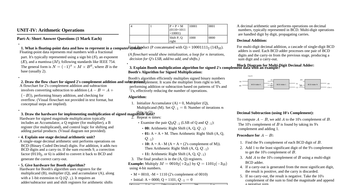
### Circulatory System: Overview The circulatory system, also known as the cardiovascular system, is a vital organ system that ensures the transport of nutrients, oxygen, hormones, and waste products throughout the body. It consists of the heart, blood vessels (arteries, veins, capillaries), and blood. #### Functions: - **Transport of Gases:** Delivers oxygen from the lungs to tissues and carbon dioxide from tissues to the lungs. - **Nutrient Transport:** Carries absorbed nutrients from the digestive tract to cells. - **Waste Removal:** Transports metabolic waste products to the kidneys and liver for excretion. - **Hormone Transport:** Distributes hormones from endocrine glands to target organs. - **Immunity:** Contains immune cells and antibodies for defense against pathogens. - **Thermoregulation:** Helps regulate body temperature. ### Heart: Anatomy & Function The heart is a muscular organ located in the mediastinum, responsible for pumping blood. #### Chambers: - **Atria (2):** Upper chambers that receive blood. - **Right Atrium:** Receives deoxygenated blood from the body via superior and inferior vena cava. - **Left Atrium:** Receives oxygenated blood from the lungs via pulmonary veins. - **Ventricles (2):** Lower chambers that pump blood out. - **Right Ventricle:** Pumps deoxygenated blood to the lungs via the pulmonary artery. - **Left Ventricle:** Pumps oxygenated blood to the body via the aorta (strongest chamber). #### Valves: - **Atrioventricular (AV) Valves:** Prevent backflow into atria. - **Tricuspid Valve:** Between right atrium and right ventricle. - **Mitral (Bicuspid) Valve:** Between left atrium and left ventricle. - **Semilunar Valves:** Prevent backflow into ventricles. - **Pulmonary Valve:** Between right ventricle and pulmonary artery. - **Aortic Valve:** Between left ventricle and aorta. #### Blood Flow Through the Heart: 1. Deoxygenated blood enters **Right Atrium** from vena cava. 2. Passes through **Tricuspid Valve** to **Right Ventricle**. 3. Pumped through **Pulmonary Valve** into **Pulmonary Artery** to lungs. 4. Oxygenated blood returns from lungs via **Pulmonary Veins** to **Left Atrium**. 5. Passes through **Mitral Valve** to **Left Ventricle**. 6. Pumped through **Aortic Valve** into **Aorta** to systemic circulation. ### Blood Vessels #### Arteries: - Carry oxygenated blood away from the heart (except pulmonary artery). - Thick, muscular, elastic walls to withstand high pressure. - Branch into smaller arterioles. #### Capillaries: - Microscopic vessels forming networks within tissues. - Site of exchange of gases, nutrients, and waste products between blood and cells. - Single-cell thick walls for efficient diffusion. #### Veins: - Carry deoxygenated blood back to the heart (except pulmonary veins). - Thinner, less muscular walls than arteries. - Contain valves to prevent backflow of blood, especially in limbs. - Merge from venules into larger veins. ### Foetal Circulatory System: Specialized Adaptations The fetal circulatory system has unique features to bypass the non-functional fetal lungs and liver, relying on the placenta for gas and nutrient exchange. #### Key Differences from Adult Circulation: - **Placenta:** Site of gas, nutrient, and waste exchange with maternal blood. - **Umbilical Cord:** Contains: - **Two Umbilical Arteries:** Carry deoxygenated blood and waste from fetus to placenta. - **One Umbilical Vein:** Carries oxygenated blood and nutrients from placenta to fetus. - **Pulmonary Bypass:** Lungs are not functional for gas exchange. - **Hepatic Bypass:** Liver is partially bypassed. #### Foetal Shunts: 1. **Ductus Venosus:** - **Location:** Connects umbilical vein directly to the inferior vena cava. - **Function:** Bypasses the fetal liver, allowing most oxygenated blood from the placenta to go directly to the heart. - **Fate after birth:** Closes to become the Ligamentum Venosum. 2. **Foramen Ovale:** - **Location:** An opening in the interatrial septum between the right and left atria. - **Function:** Allows oxygenated blood from the right atrium (coming from the placenta via ductus venosus) to bypass the pulmonary circulation and enter the left atrium, then to the left ventricle and aorta. - **Fate after birth:** Closes to become the Fossa Ovalis. 3. **Ductus Arteriosus:** - **Location:** Connects the pulmonary artery to the aorta. - **Function:** Shunts most of the deoxygenated blood from the right ventricle (which would otherwise go to the lungs) directly into the aorta, bypassing the pulmonary circulation. - **Fate after birth:** Closes to become the Ligamentum Arteriosum. #### Blood Flow in Foetus: 1. Oxygenated blood from placenta via **Umbilical Vein**. 2. Most blood bypasses liver via **Ductus Venosus** into Inferior Vena Cava. 3. Mixes with deoxygenated blood in IVC, enters **Right Atrium**. 4. Most blood passes through **Foramen Ovale** to Left Atrium. 5. From Left Atrium to Left Ventricle, then pumped to Aorta for systemic circulation. 6. Some blood from Right Atrium goes to Right Ventricle, then to Pulmonary Artery. 7. Most blood in Pulmonary Artery bypasses lungs via **Ductus Arteriosus** into Aorta. 8. Deoxygenated blood and waste products return to placenta via **Umbilical Arteries**. ### Changes at Birth Significant changes occur at birth as the baby takes its first breath and the placenta is no longer needed. - **First breath:** Lungs inflate, pulmonary vascular resistance decreases dramatically. - **Clamping of umbilical cord:** Stops blood flow from placenta. - Umbilical arteries and vein constrict. - Ductus venosus closes. - **Increased left atrial pressure:** Due to increased pulmonary blood flow, pressure in the left atrium rises, closing the **Foramen Ovale**. - **Increased oxygen tension:** Leads to constriction and closure of the **Ductus Arteriosus**. These changes redirect blood flow to the lungs for gas exchange and establish the adult circulatory pattern.