Body Fluids & Circulation
Cheatsheet Content
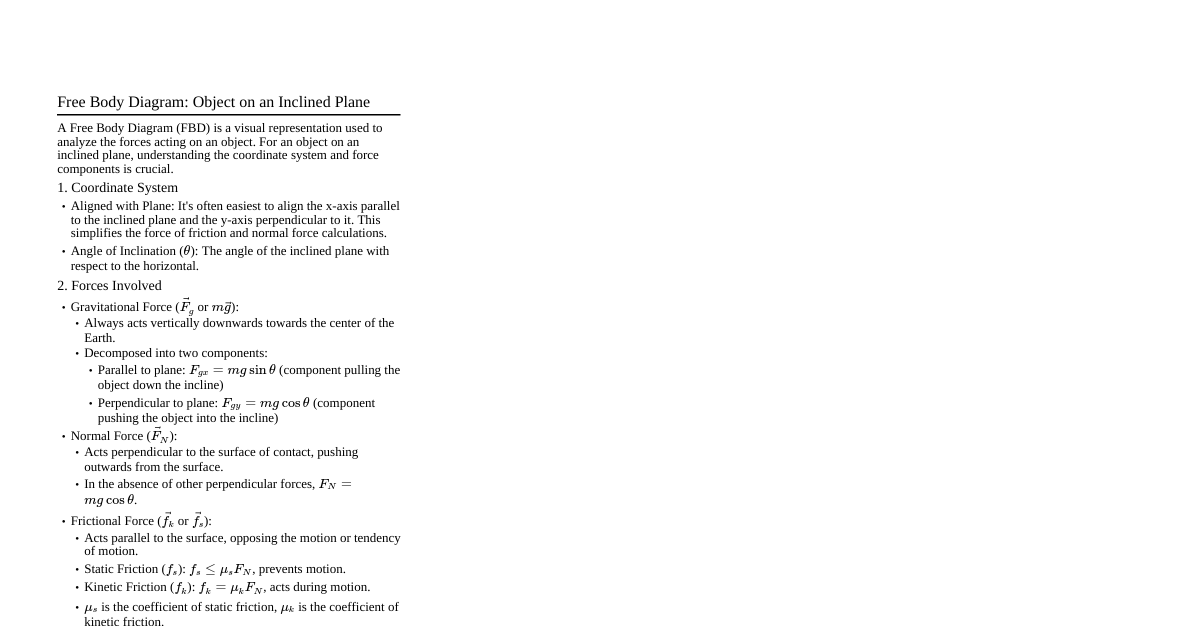
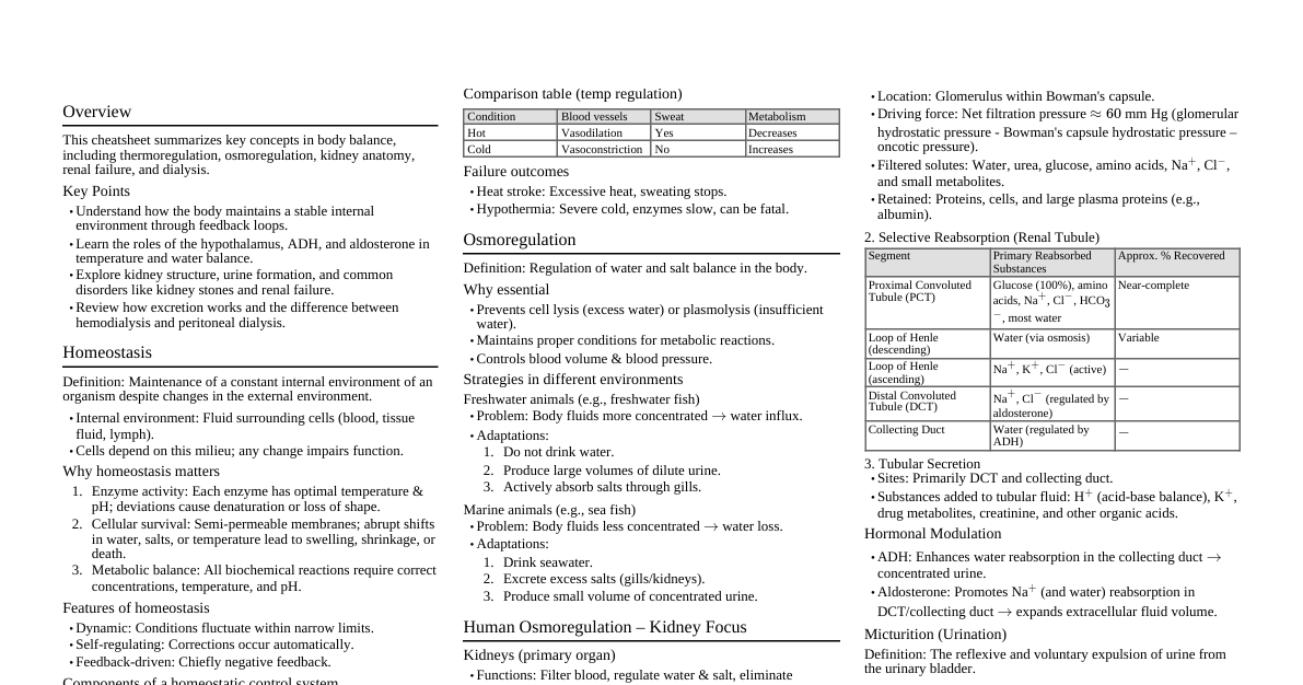
1. Introduction to Body Fluids Definition: Body fluids are the watery solutions of dissolved substances present in the body. They facilitate transport of nutrients, gases, hormones, and waste products. Types: Intracellular Fluid (ICF): Fluid within cells. (approx. 2/3 of total body water) Extracellular Fluid (ECF): Fluid outside cells. (approx. 1/3 of total body water) Interstitial Fluid (Tissue Fluid): Surrounds cells directly. Plasma: Fluid component of blood. Lymph: Fluid in lymphatic vessels. Other ECF: Cerebrospinal fluid, synovial fluid, aqueous humor, etc. 2. Blood 2.1. Composition of Blood Blood Plasma (55% of blood volume): Straw-colored, viscous fluid. Water: 90-92% Proteins (6-8%): Fibrinogen: Blood clotting. Globulins ($\alpha, \beta, \gamma$): Defense mechanisms (antibodies), transport. Albumins: Osmotic balance. Other components: Glucose, amino acids, lipids, cholesterol, mineral ions ($Na^+, Ca^{2+}, Mg^{2+}, HCO_3^-, Cl^-$), hormones, waste products. Serum: Plasma minus clotting factors (fibrinogen). Formed Elements (45% of blood volume): Produced in bone marrow. Erythrocytes (Red Blood Cells - RBCs): Most abundant (5-5.5 million/$mm^3$ in healthy adult male). Biconcave, anucleated in mammals. Contain hemoglobin (red, iron-containing protein) for $O_2$ transport. Lifespan: 120 days; destroyed in spleen ("graveyard of RBCs") and liver. Erythropoiesis: Formation of RBCs, stimulated by erythropoietin (from kidney). Leukocytes (White Blood Cells - WBCs): Fewer in number (6000-8000/$mm^3$). Nucleated, generally short-lived. Granulocytes: (contain granules) Neutrophils (60-65%): Phagocytic, first line of defense against infection. Eosinophils (2-3%): Associated with allergic reactions and parasitic infections. Basophils (0.5-1%): Secrete histamine (inflammatory response), serotonin, heparin (anticoagulant). Agranulocytes: (no granules) Lymphocytes (20-25%): B-lymphocytes: Produce antibodies (humoral immunity). T-lymphocytes: Cell-mediated immunity. Monocytes (6-8%): Phagocytic, differentiate into macrophages. Platelets (Thrombocytes): Cell fragments produced from megakaryocytes in bone marrow. 1.5-3.5 lakh/$mm^3$. Involved in blood coagulation/clotting. 2.2. Blood Groups Based on presence/absence of antigens (chemicals that induce immune response) on RBC surface and antibodies (proteins produced in response to antigens) in plasma. ABO Grouping: Blood Group Antigens on RBCs Antibodies in Plasma Can Donate To Can Receive From A A anti-B A, AB A, O B B anti-A B, AB B, O AB A, B None AB A, B, AB, O (Universal Recipient) O None anti-A, anti-B A, B, AB, O (Universal Donor) O Rh Grouping: Rh antigen (D antigen): Present in Rh-positive ($Rh^+$) individuals (approx. 80%). Absent in Rh-negative ($Rh^-$) individuals. Anti-Rh antibodies are NOT naturally present in $Rh^-$ individuals. They are produced only upon exposure to $Rh^+$ blood. Erythroblastosis foetalis: Severe hemolytic disease of the newborn. Occurs when $Rh^-$ mother carries $Rh^+$ foetus. During first pregnancy, mother's blood usually doesn't mix significantly with foetal blood, so she may develop antibodies only after delivery. In subsequent $Rh^+$ pregnancies, maternal anti-Rh antibodies cross placenta, destroy foetal RBCs, leading to severe anemia and jaundice. Prevention: Administer anti-Rh antibodies to mother immediately after delivery of first $Rh^+$ baby to destroy any foetal RBCs in her circulation. 2.3. Coagulation of Blood (Blood Clotting) Mechanism to prevent excessive blood loss. Process: Injury to blood vessel triggers release of clotting factors (thromboplastin) from damaged tissues and platelets. Clotting factors, in presence of $Ca^{2+}$, activate prothrombin activator complex. Prothrombin activator converts inactive prothrombin (plasma protein) to active thrombin . Thrombin converts soluble fibrinogen (plasma protein) into insoluble fibrin monomers. Fibrin monomers polymerize to form a network of threads. Fibrin threads trap RBCs and platelets to form the clot (thrombus). Key components: Platelets, Calcium ions ($Ca^{2+}$), Vitamin K (for synthesis of clotting factors). 3. Lymph (Tissue Fluid) Formation: As blood flows through capillaries, water, small soluble substances (nutrients, gases, hormones) filter out of capillaries into interstitial spaces, forming tissue fluid (interstitial fluid). Composition: Similar to plasma but lacks large proteins and RBCs. Contains WBCs (lymphocytes). Function: Medium for exchange between blood and cells. Drains into lymphatic capillaries, forming lymph. Carries absorbed fat from intestine (lacteals). Transports lymphocytes and antibodies for immune response. Returns excess tissue fluid and proteins to blood circulation. Lymphatic System: Network of lymphatic vessels, lymph nodes, and lymphoid organs (spleen, tonsils, thymus). Collects lymph and returns it to major veins. 4. Circulatory Pathways Open Circulatory System: Blood flows through open spaces (sinuses/lacunae) and directly bathes tissues. Found in arthropods and molluscs. Closed Circulatory System: Blood flows within a network of closed vessels. Found in annelids and chordates. More efficient. 5. Human Circulatory System Also called the Blood Vascular System . Consists of a muscular pumping heart, a network of blood vessels, and blood. 5.1. Heart Location: Thoracic cavity, between lungs, slightly tilted to the left. Protection: Double-walled membranous bag, pericardium , enclosing pericardial fluid. Chambers: Four chambers – two upper, smaller atria (auricles) and two lower, larger ventricles . Atria separated by inter-atrial septum. Ventricles separated by inter-ventricular septum. Atrium and ventricle of the same side separated by atrio-ventricular septum. Valves: Prevent backflow of blood. Tricuspid valve: Between right atrium and right ventricle. (3 cusps) Bicuspid (Mitral) valve: Between left atrium and left ventricle. (2 cusps) Semilunar valves: At the origin of pulmonary artery (from right ventricle) and aorta (from left ventricle). 5.2. Conduction of Heartbeat (Cardiac Cycle) Heart is myogenic (generates its own impulse). Nodes: Sinoatrial Node (SAN): Pacemaker of the heart. Located in upper right corner of right atrium. Generates impulse (70-75/min). Atrioventricular Node (AVN): Located in lower left corner of right atrium. Delays impulse slightly to allow atrial contraction. Conduction Pathway: SAN $\rightarrow$ Atrial muscles $\rightarrow$ AVN $\rightarrow$ Bundle of His (AV bundle) $\rightarrow$ Purkinje fibers $\rightarrow$ Ventricular muscles. Cardiac Cycle: Sequential event in the heart which is cyclically repeated. (approx. 0.8 seconds duration). Joint Diastole: All four chambers are relaxed, blood flows from atria to ventricles. Atrial Systole: Atria contract, pushing remaining blood into ventricles. Ventricular Systole: Ventricles contract, closing AV valves (Lub sound), opening semilunar valves, pumping blood into aorta and pulmonary artery. Ventricular Diastole: Ventricles relax, closing semilunar valves (Dub sound). Heart Sounds: 'Lub': First sound, due to closure of tricuspid and bicuspid (AV) valves at the start of ventricular systole. 'Dub': Second sound, due to closure of semilunar valves at the start of ventricular diastole. Cardiac Output: Volume of blood pumped by each ventricle per minute. Cardiac Output = Heart Rate $\times$ Stroke Volume Stroke Volume: Volume of blood pumped by each ventricle per beat (approx. 70 ml). Heart Rate: Number of beats per minute (approx. 72 beats/min). Cardiac Output $\approx 70 \text{ ml/beat} \times 72 \text{ beats/min} \approx 5 \text{ L/min}$. 5.3. Electrocardiograph (ECG) Graphical representation of the electrical activity of the heart during a cardiac cycle. P-wave: Depolarization (excitation/contraction) of atria. QRS complex: Depolarization of ventricles (atrial repolarization is masked). T-wave: Repolarization (relaxation) of ventricles. Deviations from normal ECG indicate abnormalities or disease. 5.4. Double Circulation Blood passes through the heart twice in one complete cycle. Pulmonary Circulation: Deoxygenated blood from right ventricle $\rightarrow$ Pulmonary artery $\rightarrow$ Lungs (oxygenation) $\rightarrow$ Pulmonary veins $\rightarrow$ Left atrium. Systemic Circulation: Oxygenated blood from left ventricle $\rightarrow$ Aorta $\rightarrow$ Body tissues (deoxygenation) $\rightarrow$ Vena Cavae $\rightarrow$ Right atrium. 5.5. Blood Vessels Arteries: Carry blood away from heart (usually oxygenated, except pulmonary artery). Thick, elastic walls. Veins: Carry blood towards heart (usually deoxygenated, except pulmonary veins). Thinner walls, often have valves to prevent backflow. Capillaries: Microscopic, one-cell thick vessels connect arterioles and venules. Site of exchange of materials. 6. Regulation of Cardiac Activity Heartbeat is regulated extrinsically by nervous and hormonal mechanisms. Neural Control: Medulla oblongata (cardiac centre): Regulates heart rate. Sympathetic nerves: Increase heart rate, strength of ventricular contraction, and cardiac output. Parasympathetic nerves (vagus nerve): Decrease heart rate and cardiac output. Hormonal Control: Adrenal medullary hormones (adrenaline, noradrenaline): Increase heart rate. 7. Disorders of Circulatory System High Blood Pressure (Hypertension): Normal BP: 120/80 mmHg (systolic/diastolic). Hypertension: $\ge 140/90$ mmHg. Can lead to heart disease, kidney disease, stroke. Coronary Artery Disease (CAD)/Atherosclerosis: Narrowing of lumen of coronary arteries due to deposition of fat, cholesterol, fibrous tissue, and calcium (plaque formation). Reduces blood supply to heart muscle. Angina Pectoris (Angina): Acute chest pain due to insufficient oxygen reaching heart muscle. Occurs when blood flow is restricted but not completely blocked. Heart Failure: State where the heart is not pumping blood effectively enough to meet the needs of the body. Sometimes called congestive heart failure because congestion of lungs is a major symptom. NOT the same as cardiac arrest (heart stops beating) or heart attack (heart muscle suddenly damaged by inadequate blood supply). Myocardial Infarction (Heart Attack): Death of heart muscle tissue due to complete blockage of a coronary artery.