Gametogenesis
Shared 3/23/2026•78 views
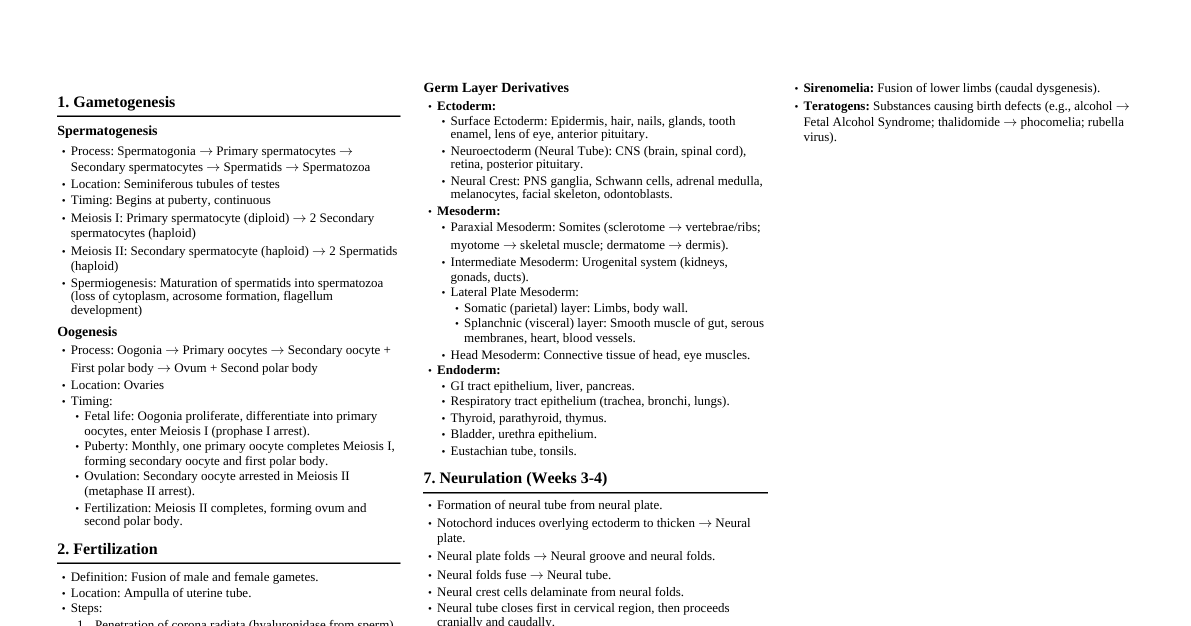
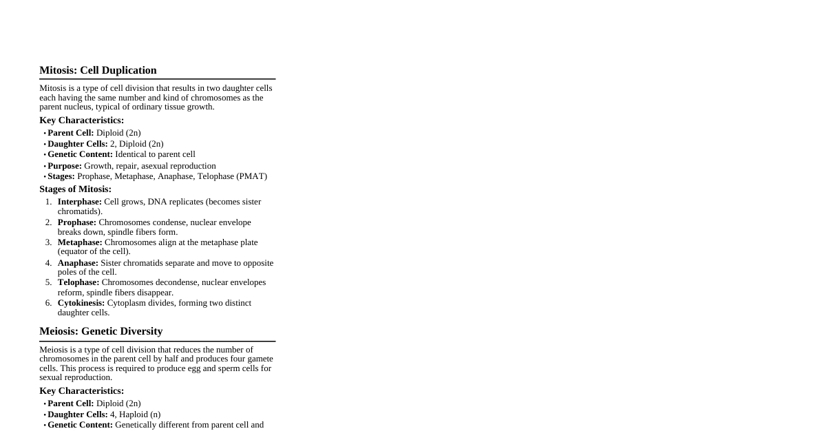
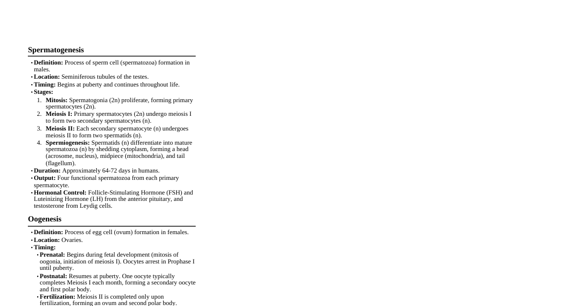
### Introduction to Gametogenesis Gametogenesis is the biological process of creating gametes (sex cells). In humans, this involves meiosis, a specialized type of cell division that reduces the chromosome number by half, producing haploid cells from diploid cells. This ensures that when two gametes fuse during fertilization, the resulting zygote has the correct diploid number of chromosomes. Key processes include: - **Meiosis:** Reductional division of chromosomes. - **Cell Differentiation:** Development of immature germ cells into mature gametes. - **Genetic Recombination:** Exchange of genetic material during meiosis, increasing genetic diversity. There are two main types of gametogenesis: 1. **Spermatogenesis:** Formation of sperm in males. 2. **Oogenesis:** Formation of eggs (ova) in females. ### Spermatogenesis: Overview Spermatogenesis is the process by which male primordial germ cells (spermatogonia) develop into mature spermatozoa. This continuous process occurs in the seminiferous tubules of the testes and takes approximately 64-72 days in humans. #### Stages of Spermatogenesis 1. **Mitotic Proliferation:** Spermatogonia (diploid stem cells) divide mitotically to produce more spermatogonia and primary spermatocytes. This ensures a continuous supply of germ cells. 2. **Meiosis I:** Primary spermatocytes (diploid, 2n) undergo Meiosis I to form two secondary spermatocytes (haploid, n, but with duplicated chromatids). 3. **Meiosis II:** Each secondary spermatocyte undergoes Meiosis II to form two spermatids (haploid, n, with single chromatids). Thus, one primary spermatocyte yields four spermatids. 4. **Spermiogenesis:** Spermatids undergo morphological changes to become mature spermatozoa (sperm). This is a differentiation process, not cell division. #### Hormonal Regulation - **GnRH (Gonadotropin-Releasing Hormone):** Released by the hypothalamus, stimulates the pituitary. - **FSH (Follicle-Stimulating Hormone):** Secreted by the anterior pituitary, stimulates Sertoli cells to support spermatogenesis and produce androgen-binding protein (ABP). - **LH (Luteinizing Hormone):** Secreted by the anterior pituitary, stimulates Leydig cells to produce testosterone. - **Testosterone:** Essential for the development and maintenance of male secondary sexual characteristics and directly stimulates spermatogenesis. - **Inhibin:** Produced by Sertoli cells, inhibits FSH release. ### Spermiogenesis: From Spermatid to Sperm Spermiogenesis is the final stage of spermatogenesis, where a round spermatid transforms into an elongated, motile spermatozoon. #### Key Morphological Changes - **Acrosome Formation:** The Golgi apparatus forms the acrosomic vesicle, which spreads over the anterior half of the nucleus to become the acrosome. The acrosome contains hydrolytic enzymes (e.g., hyaluronidase, acrosin) vital for penetrating the egg's outer layers. - **Nuclear Condensation:** The nucleus becomes highly condensed and elongated. Histones are replaced by protamines, making the DNA more compact and resistant to damage. - **Flagellum (Tail) Formation:** Centrioles migrate to the posterior pole of the nucleus. The distal centriole forms the axoneme, the core of the flagellum, which is responsible for motility. Mitochondria aggregate around the base of the flagellum to form the midpiece, providing ATP for movement. - **Cytoplasm Shedding:** Most of the spermatid's cytoplasm is shed as a residual body, which is phagocytosed by Sertoli cells. #### Structure of a Mature Spermatozoon - **Head:** Contains the haploid nucleus and the acrosome. - **Midpiece:** Contains mitochondria for energy. - **Tail (Flagellum):** Provides motility. ### Oogenesis: Overview Oogenesis is the process by which female primordial germ cells (oogonia) develop into mature ova (eggs). This process is discontinuous and begins prenatally, arresting at certain stages, and is completed only upon fertilization. #### Stages of Oogenesis 1. **Prenatal Mitotic Proliferation:** Oogonia (diploid stem cells) divide mitotically in the fetal ovary to produce primary oocytes. This proliferation stops before birth. All primary oocytes are formed by the fifth month of fetal development. 2. **Meiosis I (Arrested):** Primary oocytes (diploid, 2n) begin Meiosis I but arrest at Prophase I (specifically, in the diplotene stage) until puberty. Each primary oocyte is surrounded by follicular cells, forming a primordial follicle. 3. **Meiosis I Completion (Puberty to Menopause):** Starting at puberty, one primary oocyte per menstrual cycle (usually) completes Meiosis I, producing a large secondary oocyte (haploid, n, with duplicated chromatids) and a small first polar body. The first polar body typically degenerates. 4. **Meiosis II (Arrested):** The secondary oocyte begins Meiosis II but arrests at Metaphase II. It is then ovulated. 5. **Meiosis II Completion (Fertilization):** Meiosis II is only completed if the secondary oocyte is fertilized by a sperm. This produces a mature ovum (haploid, n, with single chromatid) and a second polar body. The second polar body also typically degenerates. #### Comparison with Spermatogenesis | Feature | Spermatogenesis | Oogenesis | | :------------------- | :---------------------------------------------- | :-------------------------------------------- | | **Location** | Testes | Ovaries | | **Onset** | Puberty | Fetal life | | **Duration** | Continuous, ~64-72 days | Discontinuous, decades | | **Gametes per Primary** | 4 functional spermatozoa | 1 functional ovum + 2-3 polar bodies | | **Size of Gametes** | Small, motile | Large, non-motile | | **Cytoplasm Division** | Equal | Unequal (polar bodies) | ### Folliculogenesis: Oocyte Development within Follicles Folliculogenesis is the maturation of the ovarian follicle, which houses and nourishes the developing oocyte. This process occurs concurrently with oogenesis. #### Stages of Follicle Development 1. **Primordial Follicle:** Primary oocyte arrested in Prophase I, surrounded by a single layer of flattened follicular cells. Present from birth. 2. **Primary Follicle:** Follicular cells become cuboidal and proliferate, forming a stratified layer (granulosa cells). A thick glycoprotein layer, the zona pellucida, forms around the oocyte. 3. **Secondary Follicle:** Granulosa cells continue to proliferate, and fluid-filled spaces appear among them, which coalesce to form the antrum. Theca cells differentiate outside the granulosa layer into theca interna (endocrine) and theca externa (connective tissue). 4. **Tertiary (Graafian) Follicle:** A large, mature follicle with a prominent antrum. The oocyte is surrounded by a mound of granulosa cells called the cumulus oophorus. This follicle is ready for ovulation. #### Hormonal Regulation of Folliculogenesis - **FSH:** Stimulates the growth of primary follicles and the proliferation of granulosa cells. - **LH Surge:** A rapid increase in LH triggers ovulation, causing the Graafian follicle to rupture and release the secondary oocyte. It also promotes the luteinization of the remaining follicular cells to form the corpus luteum. - **Estrogen:** Produced by granulosa cells (with help from theca cells), stimulates uterine lining proliferation and has feedback effects on the hypothalamus and pituitary. - **Progesterone:** Produced by the corpus luteum after ovulation, prepares the uterus for pregnancy and maintains the uterine lining. ### Fertilization and Zygote Formation Fertilization is the fusion of male and female gametes to form a zygote. #### Key Steps 1. **Sperm Capacitation:** Physiological changes in sperm within the female reproductive tract, enhancing their motility and ability to undergo the acrosome reaction. 2. **Acrosome Reaction:** Release of hydrolytic enzymes from the acrosome upon binding to the zona pellucida, allowing sperm to penetrate. 3. **Penetration of Zona Pellucida:** Sperm enzymes and tail movements create a path through the zona pellucida. 4. **Fusion of Membranes:** The sperm plasma membrane fuses with the oocyte plasma membrane. The sperm nucleus enters the oocyte cytoplasm. 5. **Cortical Reaction (Block to Polyspermy):** Upon sperm entry, cortical granules in the oocyte release enzymes that modify the zona pellucida, preventing entry of additional sperm. 6. **Meiosis II Completion:** The oocyte completes Meiosis II, forming a mature ovum and a second polar body. 7. **Pronuclei Formation:** The haploid sperm nucleus and the haploid ovum nucleus (now called pronuclei) swell and replicate their DNA. 8. **Zygote Formation:** The pronuclei fuse, restoring the diploid chromosome number and forming a zygote. #### Significance of Fertilization - Restoration of diploid chromosome number. - Determination of genetic sex (XX or XY). - Initiation of cleavage (embryonic development). - Introduction of genetic variation from two parents. ### Genetic Variation and Meiosis Gametogenesis, particularly meiosis, is crucial for introducing genetic variation. #### Mechanisms of Genetic Variation 1. **Crossing Over (Prophase I):** Homologous chromosomes exchange segments of genetic material, creating new combinations of alleles on chromatids. 2. **Independent Assortment (Metaphase I):** Homologous chromosome pairs align randomly at the metaphase plate, leading to different combinations of maternal and paternal chromosomes in the resulting gametes. 3. **Random Fertilization:** The random fusion of any sperm with any egg further increases the genetic diversity of offspring. #### Consequences of Errors in Meiosis - **Nondisjunction:** Failure of homologous chromosomes to separate during Meiosis I, or sister chromatids to separate during Meiosis II. - Results in aneuploidy (abnormal chromosome number) in gametes. - Examples: Trisomy 21 (Down Syndrome), Monosomy X (Turner Syndrome), Trisomy XXY (Klinefelter Syndrome). - **Translocations:** Exchange of segments between non-homologous chromosomes. - **Deletions/Duplications:** Loss or gain of chromosome segments. These errors can lead to developmental abnormalities, miscarriages, or genetic disorders.