Postpartum Hemorrhage (PPH)
Cheatsheet Content
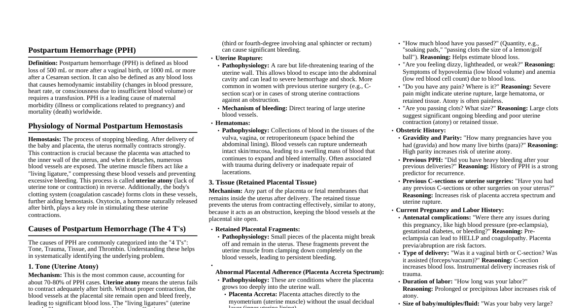
1. Definition and Significance Postpartum Hemorrhage (PPH) is defined as blood loss of 500 mL or more following a vaginal birth, or 1000 mL or more following a Cesarean section (C-section), within 24 hours of delivery ( primary PPH ). Secondary PPH is excessive bleeding occurring between 24 hours and up to 12 weeks postpartum. PPH is a leading cause of maternal mortality worldwide, meaning it is one of the most common reasons mothers die during or after childbirth. The mechanisms involve the failure of the physiological processes that normally prevent excessive bleeding after placental delivery. 2. Pathophysiology and Mechanisms of PPH (The 4 Ts) The causes of PPH are traditionally categorized into the "4 Ts": Tone, Trauma, Tissue, and Thrombin . Each 'T' represents a distinct underlying mechanism leading to excessive bleeding. 2.1. Tone (Uterine Atony) Uterine atony is the most common cause of PPH, accounting for approximately 70-80% of cases. It refers to the failure of the uterus to contract effectively after the delivery of the placenta. The normal physiological process after placental separation involves strong, sustained contractions of the myometrium (the muscular wall of the uterus). These contractions act as "living ligatures" (natural clamps) that compress the blood vessels that supplied the placenta, preventing blood loss from the open vascular sinuses (spaces filled with blood) at the placental implantation site. Mechanism: After the placenta detaches, numerous open blood vessels are exposed in the uterine wall. Normally, the uterine muscle fibers contract and retract, physically squeezing these blood vessels shut. In atony, these muscle fibers are weak or fail to contract adequately. This failure to contract allows the blood vessels to remain open and bleed freely into the uterine cavity and externally. Risk Factors and their Mechanisms: Overdistension of the Uterus: Occurs in conditions like multiple gestation (twins, triplets), polyhydramnios (excessive amniotic fluid), or a large baby (macrosomia). Mechanism: The uterine muscle fibers are stretched excessively, making them less able to contract forcefully and efficiently after delivery. Think of an overstretched rubber band that loses its snap. Prolonged Labor or Rapid Labor: Mechanism (Prolonged): The uterine muscles become fatigued from prolonged, sustained contractions, similar to any other muscle in the body. This exhaustion leads to decreased contractile strength. Mechanism (Rapid): The uterus may not have sufficient time to coordinate and establish sustained, forceful contractions after a very quick delivery. High Parity: Women who have had many previous pregnancies and deliveries. Mechanism: Repeated stretching and contracting of the uterus over multiple pregnancies can lead to a less efficient musculature, losing some of its tone and contractility over time. Chorioamnionitis: Infection of the amniotic fluid and membranes. Mechanism: Infection can cause inflammation and interfere with the normal physiological contractile response of the myometrial cells. Endotoxins from bacteria can also directly inhibit uterine contractility. Retained Placental Fragments: See "Tissue" section below for detailed mechanism. When parts of the placenta remain, the uterus cannot contract fully. Uterine Fibroids: Benign (non-cancerous) growths in the uterine wall. Mechanism: Fibroids can interfere with the normal arrangement and function of the myometrial muscle fibers, preventing uniform and effective contractions. Magnesium Sulfate Administration: Used to prevent seizures in pre-eclampsia or for fetal neuroprotection. Mechanism: Magnesium is a smooth muscle relaxant. It reduces calcium influx into muscle cells, which is essential for muscle contraction. This effect extends to the uterine musculature, decreasing its ability to contract. General Anesthesia (especially halogenated agents like halothane, isoflurane): Mechanism: These agents are potent smooth muscle relaxants, directly inhibiting uterine contractility. Placenta Previa or Abruptio Placentae: Conditions where the placenta is abnormally implanted or separates prematurely. Mechanism: These conditions can lead to a larger than normal placental implantation site or damage to the uterine wall, making it harder for the uterus to contract effectively post-delivery. 2.2. Trauma (Genital Tract Trauma) This refers to lacerations (tears) or hematomas (collections of blood outside blood vessels) in the birth canal (cervix, vagina, perineum) or uterus itself. This is the second most common cause of PPH. Mechanism: Direct injury to blood vessels during delivery results in active bleeding. Unlike uterine atony where bleeding is from the placental site, here the bleeding source is the site of the tear. The uterus may be well-contracted, but blood continues to flow from the laceration. Types and Risk Factors: Cervical Lacerations: Tears in the cervix. Risk Factors: Forceps or vacuum-assisted delivery, rapid cervical dilation, large baby. Mechanism: The cervix is highly vascular. A tear can involve significant blood vessels, leading to profuse bleeding. Vaginal or Perineal Lacerations: Tears in the vagina or perineum (the area between the vagina and anus). Risk Factors: Episiotomy (surgical cut to enlarge the vaginal opening), instrumental delivery, rapid delivery, large baby. Mechanism: Tears can extend deeply into highly vascular tissues, causing significant blood loss. Uterine Rupture: A tear in the wall of the uterus, a rare but life-threatening event. Risk Factors: Previous C-section (scar rupture is most common), prolonged labor, excessive oxytocin stimulation, high parity, uterine anomalies. Mechanism: The integrity of the uterine wall is compromised, leading to massive internal and/or external hemorrhage. Blood can escape into the abdominal cavity. Hematomas: Collections of blood, usually in the vulva, vagina, or retroperitoneum (space behind the abdominal lining). Mechanism: Blood vessels are damaged, and blood leaks into surrounding tissues, forming a localized swelling. This can mask significant blood loss, as the bleeding is not always immediately visible externally. They can expand rapidly and cause severe pain and hypovolemia (low blood volume). 2.3. Tissue (Retained Placental Tissue) This refers to the incomplete separation or expulsion of the placenta or any fragments of placental tissue remaining in the uterus after delivery. Mechanism: When placental tissue remains, the uterus cannot fully contract and retract. The retained tissue acts as a physical barrier, preventing the myometrial muscle fibers from clamping down completely on the open blood vessels at the placental implantation site. This leads to continuous bleeding from these vessels. Types and Risk Factors: Retained Placental Fragments: Small pieces of the placenta left behind. Risk Factors: Manual removal of the placenta (where parts might be missed), abnormally adherent placenta (e.g., placenta accreta). Mechanism: The presence of even small fragments prevents complete uterine contraction. Placenta Accreta Spectrum (PAS): A group of conditions where the placenta implants abnormally into the uterine wall, failing to separate normally after birth. Placenta Accreta: Chorionic villi (finger-like projections of the placenta) adhere to the myometrium without invading it. Placenta Increta: Chorionic villi invade into the myometrium. Placenta Percreta: Chorionic villi invade through the myometrium and potentially into adjacent organs (e.g., bladder). Risk Factors: Previous C-section (most significant), placenta previa, advanced maternal age, previous uterine surgery. Mechanism: The placenta is pathologically attached and cannot separate cleanly from the uterine wall. Attempts at manual removal can cause severe hemorrhage due to tearing of the uterine wall and exposed blood vessels. Succenturiate Lobe: An accessory (extra) lobe of the placenta that is connected to the main placenta by blood vessels. Mechanism: If the main placenta is expelled but the succenturiate lobe remains, it acts as retained tissue, preventing uterine contraction and causing bleeding. 2.4. Thrombin (Coagulopathy) This refers to disorders of blood clotting, where the blood's ability to form clots is impaired. This is the least common cause of PPH but can significantly exacerbate bleeding from any of the other 'T's. Mechanism: Normal hemostasis (stopping bleeding) involves primary hemostasis (platelet plug formation) and secondary hemostasis (coagulation cascade leading to fibrin clot formation). In coagulopathy, one or more components of this system are deficient or dysfunctional, leading to prolonged bleeding and inability to form stable clots. Causes and Mechanisms: Pre-existing Coagulation Disorders: Inherited: Von Willebrand disease (deficiency/defect in von Willebrand factor, important for platelet adhesion), Hemophilia (deficiency in clotting factors like Factor VIII or IX), other rare factor deficiencies. Acquired: Liver disease (liver produces most clotting factors), severe nutritional deficiencies. Mechanism: The blood lacks the necessary proteins or platelets to form a stable clot, leading to continuous bleeding. Acquired Coagulopathy in Pregnancy/PPH: Severe Preeclampsia/HELLP Syndrome: (Hemolysis, Elevated Liver enzymes, Low Platelets). Mechanism: Platelet consumption and dysfunction, liver dysfunction (affecting clotting factor production), and microangiopathic hemolytic anemia leading to red blood cell fragmentation. Disseminated Intravascular Coagulation (DIC): A life-threatening condition where the body's clotting system is abnormally activated throughout the blood vessels, leading to widespread microclot formation. This consumes clotting factors and platelets at a rapid rate, paradoxically leading to severe bleeding elsewhere. Risk Factors: Severe abruptio placentae (placental abruption), amniotic fluid embolism, severe preeclampsia, prolonged intrauterine fetal demise (dead fetus retained in uterus), severe infection (sepsis). Mechanism: The underlying condition triggers a systemic activation of coagulation. This leads to widespread formation of small clots, which consumes platelets and clotting factors. As these resources are depleted, the body loses its ability to form clots at sites of injury (like the placental bed), leading to massive hemorrhage. Simultaneously, the body's fibrinolytic system (which breaks down clots) is also activated, further contributing to bleeding. Dilutional Coagulopathy: Occurs with massive blood transfusions where large volumes of intravenous fluids and red blood cells are given without adequate replacement of clotting factors and platelets. Mechanism: The existing clotting factors and platelets in the patient's blood become "diluted" by the transfused fluids and red blood cells, reducing their concentration below effective levels. Medication-induced: Anticoagulants (e.g., heparin, warfarin, direct oral anticoagulants), antiplatelet agents (e.g., aspirin). Mechanism: These medications are designed to inhibit various parts of the coagulation cascade or platelet function, increasing the risk of bleeding. 3. Clinical Presentation (Symptoms and Signs) The clinical presentation of PPH depends on the amount and rate of blood loss. Early recognition is crucial. Symptoms (what the patient feels): Excessive Vaginal Bleeding: The most obvious symptom. May be described as soaking through pads quickly, gushing blood, or passing large clots. Weakness, Dizziness, Lightheadedness: Due to hypovolemia (reduced blood volume) and decreased brain perfusion (blood flow to the brain). Palpitations: The heart beats faster (tachycardia) to compensate for reduced blood volume and maintain cardiac output (amount of blood pumped by the heart). Shortness of Breath (Dyspnea): Due to reduced oxygen-carrying capacity of blood (anemia) and compensatory respiratory efforts. Altered Mental Status: Confusion, disorientation, loss of consciousness in severe cases, due to severe hypovolemia and cerebral hypoperfusion. Pain: May be present with uterine atony (cramping, but often less severe than labor pains), or severe localized pain with hematomas or uterine rupture. Signs (what the clinician observes): Visible Blood Loss: Often underestimated. Healthcare providers should quantify blood loss (weighing blood-soaked materials, using calibrated drapes). Uterine Atony: On abdominal palpation, the uterus feels boggy (soft and poorly contracted) and enlarged, rather than firm and well-contracted. It may be difficult to palpate the fundus (top of the uterus) due to its relaxed state. Tachycardia: Heart rate greater than 100 beats per minute, an early compensatory sign of hypovolemia. Hypotension: Low blood pressure (systolic BP less than 90 mmHg), a later and more ominous sign of significant blood loss and impending shock. Pallor: Pale skin and mucous membranes due to anemia and peripheral vasoconstriction (narrowing of blood vessels in the skin to shunt blood to vital organs). Cool, Clammy Skin: Due to peripheral vasoconstriction and activation of the sympathetic nervous system in response to shock. Prolonged Capillary Refill Time: When pressure is applied to a nail bed and released, the color normally returns quickly (less than 2 seconds). A prolonged time indicates poor peripheral perfusion. Oliguria/Anuria: Decreased or absent urine output, due to reduced renal perfusion (blood flow to the kidneys) as the body attempts to conserve fluid. This is a sign of worsening shock. Altered Mental Status: As described above, can be observed. Signs of Trauma: Visible lacerations, rapidly expanding hematoma (a tense, painful swelling that may be purplish). 4. Differential Diagnosis (What else could it be?) While the 4 Ts cover the direct causes of PPH, it's important to consider other conditions that might mimic or contribute to bleeding or related symptoms. Non-obstetric Causes of Bleeding: Vaginal Varices: Enlarged, tortuous veins in the vagina that can rupture. Cervical Polyps: Benign growths on the cervix that can bleed, especially after manipulation. Hemorrhoids: Swollen veins in the rectum/anus, can bleed. Urinary Tract Hemorrhage: Blood from the bladder or urethra. Gastrointestinal Bleeding: Blood from the stomach or intestines. Mechanism: These are not PPH but other sources of bleeding that need to be excluded. Shock from other causes: Amniotic Fluid Embolism (AFE): A rare, life-threatening condition where amniotic fluid (fluid surrounding the baby) enters the maternal bloodstream, causing an anaphylactoid reaction (severe allergic-like reaction) and cardiovascular collapse, often accompanied by DIC. Mechanism: The foreign material (amniotic fluid components) triggers a severe inflammatory and coagulopathic response, leading to acute respiratory distress, severe hypotension, and often intractable hemorrhage due to DIC. Sepsis/Septic Shock: Severe systemic infection leading to organ dysfunction and dangerously low blood pressure. Mechanism: Widespread inflammation and release of toxins can cause vasodilation (widening of blood vessels), capillary leak, and myocardial depression, leading to hypoperfusion and shock. This can also trigger DIC. Cardiogenic Shock: Failure of the heart to pump enough blood. Mechanism: Cardiomyopathy (heart muscle disease), myocardial infarction (heart attack), or severe arrhythmias (irregular heartbeats) can lead to profoundly reduced cardiac output. Neurogenic Shock: Damage to the nervous system, often spinal cord injury, leading to loss of sympathetic tone. Mechanism: Loss of sympathetic nervous system input causes widespread vasodilation and bradycardia (slow heart rate), leading to severe hypotension. Uterine Inversion: A rare but severe complication where the uterus turns inside out, either partially or completely. Mechanism: Often caused by excessive traction on the umbilical cord during placental delivery, especially if the uterus is atonic. The fundus (top of the uterus) collapses into the uterine cavity, potentially protruding through the cervix or even outside the vagina. This causes severe pain, shock (often neurogenic due to vagal stimulation, in addition to hemorrhagic), and massive bleeding. 5. History Taking (Information Gathering) A focused history helps identify risk factors and potential causes of PPH. Antepartum (Before Delivery) History: Parity: Number of previous pregnancies carried to a viable gestational age. High parity is a risk factor for atony. Previous PPH: Strongest predictor of recurrent PPH. Previous PPH indicates a predisposition or persistent risk factor. Previous C-section or Uterine Surgery: Risk factor for placenta accreta spectrum and uterine rupture. Fibroids: Risk factor for atony. Multiple Gestation/Polyhydramnios/Macrosomia: Risk factors for uterine overdistension and atony. Preeclampsia/HELLP Syndrome: Risk factor for coagulopathy (DIC). Known Coagulopathy/Bleeding Disorders: Direct risk factor for thrombin-related PPH. Medications: Anticoagulants, antiplatelet agents. Anemia in Pregnancy: Lower baseline hemoglobin means less reserve for blood loss. Intrapartum (During Delivery) History: Duration and Nature of Labor: Prolonged or rapid labor are risk factors for atony. Induction/Augmentation with Oxytocin: Excessive oxytocin can lead to uterine muscle fatigue and atony. Type of Delivery: Vaginal vs. C-section. Instrumental delivery (forceps/vacuum) increases risk of trauma. Placental Delivery: Manual removal, retained fragments, difficult separation (suggests accreta). Anesthesia Used: General anesthesia (especially halogenated agents) can cause atony. Magnesium Sulfate Administration: Risk factor for atony. Chorioamnionitis: Risk factor for atony. Postpartum (After Delivery) History (Current Event): Onset of Bleeding: Immediately after delivery (primary PPH) or later (secondary PPH). Amount and Character of Bleeding: Estimating blood loss is difficult but crucial. Ask about number of pads soaked, clots passed. Clots suggest sluggish flow or coagulopathy. Associated Symptoms: Dizziness, weakness, pain, shortness of breath, palpitations. These indicate the systemic response to blood loss. 6. Physical Examination A systematic physical examination is essential to identify the cause and assess the severity of PPH. General Assessment: Vital Signs: Heart rate, blood pressure, respiratory rate, oxygen saturation. Reasoning: Tachycardia and hypotension are key indicators of hypovolemic shock. Tachypnea (increased respiratory rate) is a compensatory mechanism. Level of Consciousness: Alert, drowsy, confused. Reasoning: Reflects cerebral perfusion. Skin Color and Temperature: Pallor, coolness, clamminess. Reasoning: Signs of peripheral vasoconstriction and poor perfusion. Capillary Refill Time: Reasoning: Indicator of peripheral perfusion. Abdominal Examination: Fundal Height and Tone: Palpate the uterine fundus. Reasoning: A boggy, poorly contracted, enlarged uterus suggests atony. A firm, well-contracted uterus points away from atony and towards trauma or tissue. Uterine Tenderness: Reasoning: Localized tenderness may suggest uterine rupture or hematoma. Abdominal Distension/Rigidity: Reasoning: May indicate internal bleeding (e.g., in uterine rupture where blood escapes into the abdominal cavity). Genital Tract Examination (Speculum and Manual Vaginal Examination): Inspection of Perineum and Vulva: Look for lacerations, hematomas. Speculum Examination: Visualize the vagina and cervix. Reasoning: Identify cervical or vaginal lacerations as a cause of trauma. If bleeding is profuse, consider removing clots to better visualize the source. Manual Vaginal Examination: Reasoning: Palpate for vaginal or cervical hematomas. Assess for retained placental fragments (can be felt as soft, irregular masses) or uterine inversion (feeling for the fundus inside the vagina or protruding). Assess uterine tone from below. Examine Placenta: If available, inspect the delivered placenta for completeness. Reasoning: Missing cotyledons (lobes of the placenta) or torn membranes with blood vessels running to the edge suggest retained placental fragments or a succenturiate lobe. 7. Investigations Investigations are crucial for confirming diagnosis, assessing severity, identifying coagulopathy, and guiding management. Immediate Bedside Tests: Visual Estimation of Blood Loss: Although notoriously inaccurate, it's the first step. Use calibrated drapes, weigh pads/swabs (1g = 1mL blood). Urine Output: Insert a Foley catheter. Reasoning: Monitor hourly urine output. Less than 30 mL/hour indicates inadequate renal perfusion and worsening shock. Laboratory Tests (STAT - as quickly as possible): Complete Blood Count (CBC): Hemoglobin (Hb) and Hematocrit (Hct): Measure the oxygen-carrying capacity of blood. Reasoning: To assess the degree of anemia and total red blood cell volume. Note that in acute bleeding, Hb/Hct may not drop immediately as compensatory fluid shifts take time. Platelet Count: Reasoning: Low platelet count (thrombocytopenia) indicates a potential coagulopathy. Coagulation Profile: Prothrombin Time (PT), Activated Partial Thromboplastin Time (aPTT), Fibrinogen, D-dimer: Reasoning: These tests assess the function of the extrinsic, intrinsic, and common pathways of the coagulation cascade, and the level of fibrinogen (a key clotting protein). Abnormal results indicate a coagulopathy (e.g., DIC, pre-existing bleeding disorder). D-dimer is elevated in DIC. Type and Crossmatch: Reasoning: To prepare for blood transfusion. Type identifies the blood group (A, B, AB, O), and crossmatch ensures compatibility with donor blood. Serum Electrolytes and Renal Function Tests (Creatinine, BUN): Reasoning: To assess for electrolyte imbalances and monitor kidney function, which can be affected by hypovolemic shock. Arterial Blood Gas (ABG) with Lactate: Reasoning: Assess acid-base status, oxygenation, and lactate levels. Elevated lactate indicates tissue hypoperfusion and anaerobic metabolism (cells producing energy without oxygen). Imaging: Ultrasound (U/S): Reasoning: Can identify retained placental fragments, assess for uterine hematomas, or in rare cases, help diagnose uterine rupture (though clinical signs are usually paramount). Can also confirm uterine atony by showing a large, flaccid uterus. CT Angiography/MRI: Reasoning: Rarely used in acute PPH due to time constraints, but may be considered for diagnosis of placenta accreta spectrum in stable patients or to locate a source of secondary PPH. 8. Management and Treatment Management of PPH is an emergency and requires a rapid, multidisciplinary approach. The primary goals are to stop the bleeding, restore blood volume, and treat the underlying cause. The general approach is often referred to as "Resuscitate and Treat". 8.1. General Principles (The "ABCDE" approach to resuscitation) Airway: Ensure patent airway. Breathing: Administer high-flow oxygen via a non-rebreather mask to optimize oxygen delivery to tissues. Circulation: Establish IV Access: Insert two large-bore (14- or 16-gauge) intravenous catheters. Reasoning: To facilitate rapid fluid and blood product administration. Fluid Resuscitation: Rapid infusion of crystalloids (e.g., normal saline or lactated Ringer's solution) while awaiting blood products. Infuse 3:1 ratio (3 mL crystalloid for every 1 mL of estimated blood loss). Mechanism: To expand intravascular volume and maintain blood pressure. Blood Transfusion: Type-specific blood, or O-negative (universal donor) blood if type-specific is not immediately available. Activate massive transfusion protocol. Mechanism: To replace lost red blood cells (for oxygen-carrying capacity), plasma (for clotting factors), and platelets. Monitor Vitals: Continuously monitor heart rate, blood pressure, respiratory rate, oxygen saturation. Foley Catheter: Insert for hourly urine output monitoring. Disability: Assess neurological status. Exposure: Keep patient warm (hypothermia can worsen coagulopathy). 8.2. Medical Management (Pharmacological) for Uterine Atony These are uterotonic agents, meaning they stimulate uterine contractions. Oxytocin (Pitocin): Mechanism: Synthetic oxytocin binds to oxytocin receptors on uterine smooth muscle cells, causing rhythmic contractions. It is the first-line uterotonic. Administration: IV infusion (e.g., 10-40 units in 500-1000 mL crystalloid, infused rapidly) or IM (intramuscular) injection (10 units). Methylergonovine (Methergine): Mechanism: An ergot alkaloid that causes strong, sustained uterine contractions (tetanic contractions) by acting on alpha-adrenergic and serotonin receptors. Administration: IM (0.2 mg). Contraindication: Hypertension (high blood pressure) or preeclampsia, as it can cause severe vasoconstriction and dangerously increase blood pressure. Carboprost Tromethamine (Hemabate): Mechanism: A synthetic prostaglandin F2-alpha analog that directly stimulates myometrial contractions and causes vasoconstriction. Administration: IM injection (250 mcg). Contraindication: Asthma (can cause bronchospasm), severe cardiac, renal, or hepatic disease. Misoprostol (Cytotec): Mechanism: A synthetic prostaglandin E1 analog that causes uterine contractions and cervical ripening. Administration: Rectal (800-1000 mcg) is common for PPH, or oral/sublingual. Advantages: Can be used in patients with hypertension or asthma. Tranexamic Acid (TXA): Mechanism: An antifibrinolytic agent that inhibits the breakdown of fibrin clots by blocking plasminogen activation. It helps stabilize existing clots. Administration: IV (1g over 10 minutes, repeated once if needed). Recommendation: Administer within 3 hours of bleeding onset for optimal benefit, based on the WOMAN trial. 8.3. Mechanical Management Uterine Massage: Mechanism: Manual compression and rubbing of the uterus stimulates contractions by directly activating muscle fibers and expelling clots. This is a first-line intervention for atony. Bimanual Compression: Mechanism: One hand is placed in the vagina, compressing the anterior uterine wall against the posterior uterine wall which is compressed by the other hand on the abdomen. This directly compresses the uterine blood vessels and stimulates contractions. Intrauterine Balloon Tamponade (e.g., Bakri balloon, Sengstaken-Blakemore tube): Mechanism: A balloon catheter is inserted into the uterus and inflated with saline. The inflated balloon exerts internal pressure on the uterine walls, mechanically compressing the bleeding vessels at the placental site. This acts as a physical barrier to bleeding. Reasoning: A temporary measure to control bleeding while other interventions are prepared or take effect. Uterine Packing: Mechanism: Packing the uterine cavity with gauze. Less commonly used now due to infection risk and potential to mask ongoing bleeding, but can be effective by applying direct pressure. 8.4. Surgical Management Repair of Lacerations: Mechanism: Direct suturing (stitching) of cervical, vaginal, or perineal tears stops bleeding by ligating (tying off) damaged blood vessels. Manual Removal of Retained Placental Fragments: Mechanism: The healthcare provider inserts a hand into the uterus to manually detach and remove any remaining placental tissue. This allows the uterus to contract fully. Curettage: Mechanism: Surgical scraping of the uterine lining (with a curette) to remove any remaining placental tissue. Often done after manual removal. Uterine Artery Embolization (UAE): Mechanism: A minimally invasive procedure performed by interventional radiologists. A catheter is inserted into the femoral artery and guided to the uterine arteries. Embolic agents (small particles) are injected to block blood flow to the uterus, thereby reducing bleeding. Reasoning: Preserves fertility. Often used when medical and other mechanical measures fail, or for secondary PPH. Surgical Ligation of Uterine or Internal Iliac Arteries: Mechanism: Open surgical procedure to tie off the main blood vessels supplying the uterus (uterine arteries) or the larger internal iliac arteries. This reduces blood flow to the uterus. Reasoning: More invasive than embolization but can be life-saving. B-Lynch Suture or other Compression Sutures: Mechanism: Specific surgical sutures applied around the uterus to mechanically compress it and stop bleeding, particularly from atony. These are fertility-sparing. Hysterectomy: Mechanism: Surgical removal of the uterus. This is a last resort, life-saving procedure when all other measures fail, especially in cases of intractable atony, severe placenta accreta spectrum, or uterine rupture. Reasoning: Removes the bleeding organ entirely. 8.5. Management of Coagulopathy Blood Component Therapy: Fresh Frozen Plasma (FFP): Contains all clotting factors. Mechanism: Replaces deficient clotting factors, especially in DIC or dilutional coagulopathy. Cryoprecipitate: Rich in fibrinogen, Factor VIII, Factor XIII, von Willebrand factor. Mechanism: Specifically used to replace fibrinogen, which is often severely depleted in DIC. Platelet Transfusion: Mechanism: Replaces platelets in cases of severe thrombocytopenia or platelet dysfunction. Specific Factor Concentrates: (e.g., Factor VIIa, prothrombin complex concentrate) Mechanism: Used in rare cases of specific factor deficiencies or refractory bleeding, to rapidly enhance clot formation. Identify and Treat Underlying Cause: (e.g., manage sepsis, deliver placenta in abruptio) Reasoning: Addressing the root cause of coagulopathy is essential for definitive treatment. 9. Secondary PPH (Bleeding > 24 hours to 12 weeks postpartum) Causes: Retained Placental Fragments (most common): Mechanism: Similar to primary PPH, retained tissue prevents full uterine involution (return to normal size) and continuous bleeding from the placental site. It can also become necrotic and infected. Endometritis: Infection of the uterine lining. Mechanism: Inflammation and infection can cause local vessel fragility, impaired clotting, and prevent proper uterine contraction, leading to bleeding. Subinvolution of the Uterus: Failure of the uterus to return to its normal non-pregnant size after childbirth. Mechanism: If the uterus remains enlarged, the blood vessels at the placental site are not adequately compressed, leading to prolonged or excessive bleeding. Can be caused by retained tissue or infection. Placental Site Subinvolution: Failure of the placental implantation site to heal properly. Mechanism: The blood vessels at the placental site remain open and prone to bleeding. Coagulopathy: Less common, but can be a factor. Management: Antibiotics: For endometritis. Uterotonics: (e.g., oral methylergonovine, oxytocin) to help uterine involution. Surgical Evacuation (D&C - Dilation and Curettage): To remove retained placental fragments. Uterine Artery Embolization: If bleeding is severe and other measures fail. 10. Complications of PPH Hypovolemic Shock: Life-threatening condition due to severe blood loss. Mechanism: Inadequate blood volume leads to insufficient oxygen delivery to tissues and organs, causing cell damage and organ failure. Acute Kidney Injury (AKI): Mechanism: Prolonged renal hypoperfusion (reduced blood flow to kidneys) leads to damage to kidney tubules (acute tubular necrosis). Adult Respiratory Distress Syndrome (ARDS): Mechanism: Severe systemic inflammation and fluid overload from massive transfusions can damage the lung capillaries, leading to fluid accumulation in the lungs and impaired gas exchange. Sheehan's Syndrome: Postpartum pituitary gland necrosis (death of pituitary cells). Mechanism: Severe hypovolemic shock can cause ischemia (reduced blood supply) to the pituitary gland, which is enlarged during pregnancy and thus more vulnerable to ischemic damage. This leads to deficiency of pituitary hormones. Disseminated Intravascular Coagulation (DIC): Can be a cause and a complication of PPH. Anemia: Chronic fatigue, weakness. Infection: Increased risk, especially with retained tissue or multiple interventions. Need for Hysterectomy: Loss of fertility. Maternal Mortality: Death of the mother.