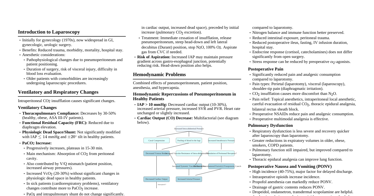
Scleral Buckling Surgery
Cheatsheet Content
### Introduction to Scleral Buckling Scleral buckling is a surgical procedure used to repair retinal detachment. It involves placing a silicone band (buckle) around the sclera (white outer layer of the eye) to indent it, bringing the choroid and retinal pigment epithelium closer to the detached retina, thereby closing the retinal breaks and allowing the retina to reattach. ### Indications for Surgery - **Rhegmatogenous Retinal Detachment (RRD):** Most common indication, caused by a break in the retina allowing fluid to pass into the subretinal space. - **Detachment with clear media:** When the view of the retina is not obscured by cataract or vitreous hemorrhage. - **Inferior or peripheral retinal breaks:** Often well-suited for buckling. - **Younger patients:** Generally preferred in younger, phakic (lens present) patients due to lower risk of cataract progression compared to vitrectomy. ### Preoperative Assessment - **Detailed history and ophthalmic examination:** Including visual acuity, intraocular pressure, and comprehensive fundus examination to identify all retinal breaks. - **B-scan ultrasonography:** If media opacities prevent direct visualization of the retina. - **Patient counseling:** Discussing risks, benefits, and expected outcomes. ### Surgical Steps The procedure typically involves several key stages: #### 1. Anesthesia - **Local or general anesthesia:** Chosen based on patient's health and surgeon's preference. #### 2. Conjunctival Incision & Muscle Isolation - A 360-degree peritomy (incision around the cornea) is created to access the sclera. - Rectus muscles are isolated with sutures to allow rotation of the globe for better access. #### 3. Localization of Retinal Breaks - The surgeon uses an indirect ophthalmoscope and scleral depressor to precisely locate all retinal breaks. - Markings are made on the sclera corresponding to the breaks. #### 4. Cryopexy or Laser Photocoagulation - **Cryopexy:** A freezing probe is applied to the outer surface of the sclera over the retinal breaks. This causes a chorioretinal adhesion, sealing the breaks. - **Laser Photocoagulation:** Less common during the primary buckling procedure, but can be used post-operatively or in conjunction. #### 5. Drainage of Subretinal Fluid (SRF) - Optional - If the retinal detachment is extensive or significantly elevated, SRF drainage may be performed. - A small incision is made in the sclera and choroid, allowing fluid to escape. This helps the retina settle. #### 6. Placement of Scleral Buckle - A silicone explant (solid or sponge) is selected based on the number, location, and extent of retinal breaks. - The explant is sutured to the sclera, creating an indentation. For circumferential detachments, a band may be placed around the entire globe. - The tension of the buckle is adjusted to ensure adequate indentation and closure of the breaks without causing excessive intraocular pressure. #### 7. Intraocular Gas or Oil (Rarely) - In complex cases, intraocular gas (e.g., SF6, C3F8) or silicone oil may be injected into the vitreous cavity to provide additional tamponade and flatten the retina. This is more common with vitrectomy, but can be used in conjunction with buckling. #### 8. Closure - The rectus muscles are reapproximated. - The conjunctiva is closed with absorbable sutures. ### Postoperative Care - **Eye patching and shield:** To protect the eye. - **Topical antibiotics and steroids:** To prevent infection and reduce inflammation. - **Activity restrictions:** Avoiding strenuous activities and head movements. - **Regular follow-up:** To monitor retinal reattachment and visual recovery. ### Potential Complications - **Recurrence of detachment:** Requires further intervention. - **Diplopia (double vision):** Due to muscle manipulation. - **Infection:** Rare but serious. - **Extrusion or migration of the buckle:** May require removal. - **Myopia shift:** Due to the change in eye shape from the buckle. - **Vitreous hemorrhage.** - **Cataract progression (if phakic).**